
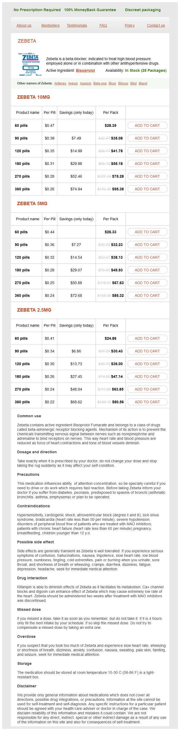
Bisoprolol Fumarate dosages: 10 mg, 5 mg
Bisoprolol Fumarate packs: 60 pills, 90 pills, 120 pills, 180 pills, 270 pills, 360 pills
In stock: 835
Only $0.24 per item
Fibers from spinal cord levels T1 to T5 pass predominantly superiorly hypertension teaching for patients discount bisoprolol 10mg fast delivery, whereas bers from T5 to L2 pass inferiorly. All sympathetics passing into the head have preganglionic bers that emerge from spinal cord level T1 and ascend in the sympathetic trunks to the highest ganglion in the neck (the superior cervical ganglion), where they synapse. Postganglionic bers then travel along blood vessels to target tissues in the head, including blood vessels, sweat glands, small smooth muscles associated with the upper eyelids, and the dilator of the pupil. Sympathetic innervation of thoracic and cervical viscera the splanchnic nerves generally connect with sympathetic ganglia around the roots of major arteries that branch from the abdominal aorta. Postganglionic sympathetic bers are distributed in extensions of this plexus, predominantly along arteries, to viscera in the abdomen and pelvis. Some of the preganglionic bers in the prevertebral plexus do not synapse in the sympathetic ganglia of the plexus, but pass through the system to the adrenal gland where they synapse directly with cells of the adrenal medulla. These cells are homologues of sympathetic postganglionic neurons and secrete adrenaline and noradrenaline into the vascular system. Preganglionic sympathetic bers may synapse with postganglionic motor neurons in ganglia and then leave the ganglia medially to innervate thoracic or cervical viscera. They may ascend in the trunk before synapsing; after synapsing, the postganglionic bers may combine with those from other levels to form named visceral nerves, such as cardiac nerves. Often, these nerves join branches from the parasympathetic system to form plexuses on or near the surface of the target organ; for example, the cardiac and pulmonary plexuses. Spinal cord levels T1 to T5 mainly innervate cranial, cervical, and thoracic viscera. The preganglionic bers in these nerves are derived from spinal cord levels T5 to L2. Ce rvic al Sympathetic cardiac nerves Sympathetic trunk Gray ramus communicans T1 to T4 Cardiac plexus Sympathetic cardiac nerves White ramus communicans 26. Like the visceral motor nerves of the sympathetic part, the visceral motor nerves of the parasympathetic part generally have two neurons in the pathway. Cranial nerve preganglionic parasympathetic bers Sacral preganglionic parasympathetic bers In the sacral region, the preganglionic parasympathetic bers form special visceral nerves (the pelvic splanchnic nerves), which originate from the anterior rami of S2 to S4 and enter pelvic extensions of the large prevertebral plexus formed around the abdominal aorta. These bers are distributed to pelvic and abdominal viscera mainly along blood vessels. Postganglionic bers leave the ganglia, join the branches of the trigeminal nerve [V], and are carried to target tissues (salivary, mucous, and lacrimal glands; constrictor muscle of the pupil; and ciliary muscle in the eye) with these branches. These branches contribute to plexuses associated with thoracic viscera or to the large prevertebral plexus in the abdomen and pelvis.
Hydrolyzed Spleen Extract (Spleen Extract). Bisoprolol Fumarate.
Source: http://www.rxlist.com/script/main/art.asp?articlekey=96976
As it passes through this part of the lesser omentum prehypertension 34 weeks pregnant generic bisoprolol 10 mg, it is anterior to the omental foramen and posterior to both the bile duct, which is slightly to its right, and the hepatic artery proper, which is slightly to its left. On approaching the liver, the portal vein divides into right and left branches, which enter the liver parenchyma. Splenic vein the splenic vein forms from numerous smaller vessels leaving the hilum of the spleen. It passes to the right, passing through the splenorenal ligament with the splenic artery and the tail of pancreas. Continuing to the right, the large, straight splenic vein is in contact with the body of the pancreas as it crosses the posterior abdominal wall. Posterior to the neck of the pancreas, the splenic vein joins the superior mesenteric vein to form the portal vein. Tributaries to the splenic vein include: short gastric veins from the fundus and left part of the greater curvature of the stomach, the left gastro-omental vein from the greater curvature of the stomach, pancreatic veins draining the body and tail of pancreas, and usually the inferior mesenteric vein. It begins in the right iliac fossa as veins draining the terminal ileum, cecum, and appendix join, and ascends in the mesentery to the right of the superior mesenteric artery. Posterior to the neck of the pancreas, the superior mesenteric vein joins the splenic vein to form the portal vein. As a corresponding vein accompanies each branch of the superior mesenteric artery, tributaries to the superior mesenteric vein include jejunal, ileal, ileocolic, right colic, and middle colic veins. Additional tributaries include: the right gastro-omental vein, draining the right part of the greater curvature of the stomach; and the anterior and posterior inferior pancreaticoduodenal veins, which pass alongside the arteries of the same name; the anterior superior pancreaticoduodenal vein usually empties into the right gastro-omental vein, and the posterior superior pancreaticoduodenal vein usually empties directly into the portal vein. Inferior mesenteric vein Superior mesenteric vein the superior mesenteric vein drains blood from the small intestine, cecum, ascending colon, and transverse colon the inferior mesenteric vein drains blood from the rectum, sigmoid colon, descending colon, and splenic exure. It begins as the superior rectal vein and ascends, receiving tributaries from the sigmoid veins and the left colic vein. All these veins accompany 179 Abdomen Liver Stomach Short gas tric veins Spleen Left gas tric vein Left gas troomental vein Portal vein Superior mes enteric vein As cending colon Splenic vein Inferior mes enteric vein Des cending colon Clinical app Hepatic cirrhosis Cirrhosis is a complex disorder of the liver, the diagnosis of which is con rmed histologically. Cirrhosis is characterized by widespread hepatic brosis interspersed with areas of nodular regeneration and abnormal reconstruction of pre-existing lobular architecture. The poorly functioning liver cells (hepatocytes) are unable to break down blood and blood products, leading to an increase in the serum bilirubin level, which manifests as jaundice. As the cirrhosis progresses, the intrahepatic vasculature is distorted, which in turn leads to increased pressure in the portal vein and its draining tributaries (portal hypertension). Portal hypertension produces increased pressure in the splenic venules leading to splenic enlargement. These veins are susceptible to bleeding and may produce marked blood loss, which in some instances can be fatal. Clinical app Portosystemic anastomosis the hepatic portal system drains blood from the visceral organs of the abdomen to the liver.
The principles outlined here for T-B cell collaboration help to explain a phenomenon that is known as the hapten-carrier effect hypertension stage 1 buy bisoprolol 10 mg without a prescription. Haptens, such as dinitrophenol, are small chemicals that can be recognized by specific antibodies but are not immunogenic by themselves. If, however, haptens are coupled to proteins, which serve as carriers, the conjugates are able to induce antibody responses against the haptens. Analysis of antibody responses to hapten-carrier conjugates provided among the earliest demonstrations of how antigen presentation by B lymphocytes contributes to the development of humoral immune responses. There are three important characteristics of anti-hapten antibody responses to hapten-protein conjugates. First, such responses require both hapten-specific B cells and protein (carrier)-specific helper T cells. Second, to stimulate a response, the hapten and carrier portions have to be physically linked and cannot be administered separately. In responses to hapten-carrier conjugates, the hapten (the B cell epitope) is recognized by a specific B cell, the conjugate is endocytosed, the carrier protein is processed in the B cell, and peptides from the carrier (the T cell epitopes) are presented to the helper T cell. Helper T Cell-Dependent Antibody Responses to Protein Antigens 259 molecules that are identical to those that were involved in the initial activation of naive T cells by dendritic cells. All of these features of antibody responses to haptenprotein conjugates can be explained by the antigenpresenting functions of B lymphocytes. Hapten-specific B cells bind the antigen through the hapten determinant, endocytose the hapten-carrier conjugate, digest the protein component, and present peptides derived from the carrier protein to carrier-specific helper T lymphocytes. Thus, the two cooperating lymphocytes recognize different epitopes of the same antigen. The hapten is responsible for efficient internalization of the carrier protein into the B cell, which explains why hapten and carrier must be physically linked. The hapten-carrier effect is the basis for the development of conjugate vaccines against encapsulated bacteria; these vaccines contain carbohydrate epitopes recognized by B cells attached to proteins recognized by T cells, discussed later in this chapter. T cell-derived cytokines are essential for germinal center reactions, described later. Several cytokines have also been implicated in the early steps of B cell proliferation and differentiation, but it is not clear if any are actually essential for these responses. After the initial interaction of B cells with helper T cells at the interface between the follicle and the T cell zone, subsequent activation of B cells by helper T cells can occur at two different locations, one outside the follicles in an extrafollicular focus and the other in the germinal centers of follicles. The antibody-secreting cells that are generated in extrafollicular foci, including plasmablasts and tissue plasma cells, are mostly short-lived, and these cells do not acquire the ability to migrate to distant sites, such as the bone marrow.
Syndromes
Additional information:
Usage: b.i.d.
Tags: bisoprolol 10 mg mastercard, quality 10 mg bisoprolol, bisoprolol 5mg overnight delivery, cheap bisoprolol 10 mg without a prescription
Snorre, 57 years: The bronchial veins drain into: either the pulmonary veins or the left atrium; and into the azygos vein on the right or into the superior intercostal vein or hemiazygos vein on the left.
Jaffar, 45 years: The number of acini per terminal duct is more than double the normal number found in normal lobules.
Lukar, 63 years: It is positioned in the midline along the posterior border of the perineal Table 5.
Peratur, 64 years: The crystals activate complement and attract neutrophils that phagocytize the crystals and then release leukotrienes, prostaglandins, free radicals, and lysosomal enzymes to produce inflammation.
C-585, Saraswati Vihar, Pitampura, New Delhi 110034


