Bromhexine
Bromhexine
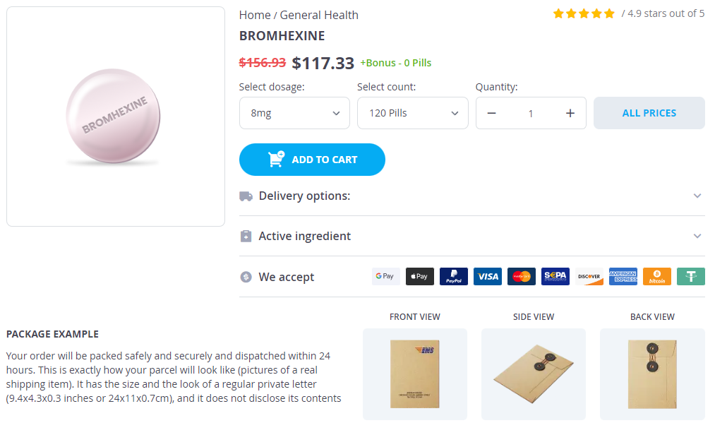
Bromhexine dosages: 8 mg
Bromhexine packs: 30 pills, 60 pills, 120 pills, 240 pills, 300 pills
In stock: 748
Only $0.91 per item
Description
An inner layer of cuboidal or columnar epithelial cells and an outer layer of myoepithelial cells line all ducts and ductules symptoms 3 days after embryo transfer discount 8mg bromhexine fast delivery. The connective tissue within the lobule is a hormonally responsive mixture of fibroblasts, occasional lymphocytes, and histiocytes, in a background of collagen and acid mucin. During pregnancy there is a marked proliferation of ductules, which results in very large lobules, and the epithelial cells have abundant cytoplasm filled with secretory vacuoles. These secretory changes usually disappear after lactation; in some cases, however, they persist for years after pregnancy and are sometimes seen even in patients who have not been pregnant. Nonproliferative lesions yield a scant specimen when the lesion is predominantly fibrous. When a cyst is present, the specimen is a fluid that may be thin and yellow or thick and darker in color. The nucleus is centrally located and round, with a prominent nucleolus, and moderate anisonucleosis is present in some cases. It has been suggested that symptomatic complicated cysts, cystic lesions with thick indistinct walls and/or thick septations, intracystic masses, and predominantly solid masses with cystic degeneration are more likely to be malignant and thus merit cytologic or histopathologic examination. The cells of this tumor, thought to be of Schwann cell origin, have a very low nuclear-to-cytoplasmic ratio, a small nucleus, and a coarsely granular cytoplasm. An apocrine carcinoma should be suspected when there is hypercellularity, marked nuclear atypia, mitotic activity, and pronounced cell dyshesion, but a cautious diagnostic approach is advisable because marked variation in nuclear size is also seen in apocrine metaplasia. Apocrine adenosis, although rare, can also display nuclear atypia, but there is less nuclear hyperchromasia than with carcinoma, and there are many naked nuclei. The spectrum includes proliferative lesions without atypia ("usual ductal hyperplasia"), atypical ductal hyperplasia, and atypical lobular hyperplasia. The criteria that define these entities and distinguish them from carcinoma in situ are histologic, not cytologic. Lesions reported cytologically as "proliferative lesion with atypia" are associated with a significantly higher frequency of histologically confirmed malignancy than those reported as "proliferative lesion without atypia" (36. They are characterized by any combination of small or large cysts, apocrine metaplasia, focal fibrosis, adenosis, and ductal hyperplasia. Cysts arise from the terminal duct lobular units by an unfolding and simplification of adjacent ductules. Moderate and florid ductal hyperplasia, which is seen in some but not all cases, is a marker for increased cancer risk. For this reason, fibrocystic changes are usually categorized as nonproliferative or proliferative, depending on whether ductal hyperplasia is present.
Oil of Thyme (Thyme). Bromhexine.
- What is Thyme?
- Dosing considerations for Thyme.
- Are there any interactions with medications?
- Bronchitis, in combination with cowslip; treating hair loss (alopecia areata) when combined with other herbs; improving movement disorders in children when used with other medicines; colic; ear infections; swelling (inflammation) of the tonsils; preventing bedwetting; sore throat; bad breath; bronchitis; and swelling (inflammation) of the lungs and mouth.
- How does Thyme work?
- Are there safety concerns?
Source: http://www.rxlist.com/script/main/art.asp?articlekey=96799
The point of bifurcation treatment nerve damage bromhexine 8 mg order overnight delivery, called the carina, is marked in the inside of the airway by a cartilaginous wedge that projects upward into the airway lumen. Courses more vertically and is shorter and wider than the left primary bronchus; gives rise to the superior, middle, and inferior secondary (lobar) bronchi. Courses more obliquely and is longer and thinner than the right primary bronchus; gives rise to the superior and inferior secondary (lobar) bronchi. Each secondary bronchus further divides into tertiary (segmental) bronchi and then continue to branch. The smallest bronchi give rise to bronchioles, which terminate in alveolar sacs, where the exchange of gases takes place. Because of its wider, more vertical orientation, the right bronchus usually has inhaled foreign objects fall into it from the trachea. Note that the esophageal hiatus is fonned by the right crus of the diaphragm, splitting to wrap around the esophagus to become the so-called esophageal sphincter. The esophagus receives its arterial supply via esophageal, left gastric, and inferior phrenic arteries. Blood drains from the distal end of the esophagus (near the diaphragm) through the (1) azygos system of veins and (2) left gastric vein, which ultimately drains into the hepatic portal vein (this knowledge will assist in understanding the complications of portal hypertension; see Chapter 10). Descends into the thoracic cavity anterior to the right subclavian artery and gives rise to the following: · Right recurrent laryngeal nerve. Hooks around the right subclavian artery and ascends back into the neck en route to intrinsic laryngeal muscles. The right vagus nerve continues to the deep surface of the esophagus, becoming the posterior vagal trunk. The right vagus nerve contributes to the cardiac plexus, which slows heart rate, and pulmonary plexus, which causes bronchoconstriction. Enters the thorax between the left common carotid and subclavian arteries and gives rise to the following: · Left recurrent laryngeal nerve. Hooks around the aortic arch by the ligamentum arteriosum and ascends back into the neck en route to the intrinsic laryngeal muscles. The left vagus nerve continues to the anterior surface of the esophagus, becoming the anterior vagal trunk. The left vagus nerve contributes to the cardiac plexus, which slows heart rate, and pulmonary plexus, which causes bronchoconstriction. The anterior and posterior vagal trunks exchange fibers, creating an esophageal plexus of nerves. The vagus nerve carries visceral sensory neurons whose sensory cell bodies are located in the inferior vagal ganglion. The visceral afferents from the vagus nerves transmit information to the brain about normal physiologic processes and visceral reflexes.
Specifications/Details
It is hypothesized that the brain cannot distinguish between sensory input from the ischemic heart tissue (as relayed by visceral sensory neurons) and cutaneous sensation (as relayed by Tl-T4 spinal nerves) administering medications 8th edition 8mg bromhexine for sale. Therefore, the patient may perceive ischemic heart pain as coming from the chest or upper limb (where the Tl-T4 dermatomes reside). The superior mediastinum is the region above the sternal angle and contains the following structures: · Aortic arch. The aortic arch arises at the level of the transverse thoracic plane, ascends up into the superior mediastinum, and descends in the posterior mediastinum. The aortic arch gives rise to the following three primary branches: · Brachiocephalic trunk. The trachea bifurcates at the level of the transverse thoracic plane into the left and right primary bronchi. The esophagus is a vertical, muscular tube that is located posterior to the trachea and transports food from the pharynx to the stomach. The phrenic nerves (en route to the diaphragm) and vagus nerves (en route to thoracic and abdominal organs) course through the superior mediastinum. The mediastinum is the anatomic region medial to the pleural sacs, between the sternum, vertebral column, rib 1, and the diaphragm. The inferior mediastinum is classically subdivided into anterior, middle, and posterior parts. Therefore, the four subregions of the mediastinum are the anterior mediastinum, middle mediastinum, posterior mediastinum, and superior mediastinum. The anterior mediastinum is deep to the sternum and bounded by the sternal angle, pericardia! The thymus in adults is involuted and is primarily a connective tissue remnant Middle mediastinum. The posterior mediastinum contains the following anatomic structures, which are posterior to the pericardia! The thoracic portion of the aorta that gives rise to posterior intercostal arteries. The primary lymphatic duct that receives lymph from all tissues below the diaphragm and from the left side of the head, neck, upper limb, and thorax. The ganglia that comprise the sympathetic chain are neuronal cell bodies from postganglionic sympathetic neurons. The ganglia associated with the sympathetic chain are organized in a vertical fashion and are segmentally linked with each ofthe thoracic spinal nerves via white and gray communicantes. The sympathetic chain is associated with the preaortic ganglia by way of greater, lesser, and least splanchnicnerves.
Syndromes
- Injury to nerves at the base of the neck called the brachial plexus
- How long does it last -- for how many day(s)?
- Meat
- Dystonia
- Nausea
- You have sudden, severe hearing loss or ringing in the ears (tinnitus)
- Parathyroid adenoma
- Culture of skin fibroblasts
- Toes that turn purple and occur with foot pain
- The immune system attacking the thyroid gland
Related Products
Additional information:
Usage: q.h.
Tags: discount bromhexine 8 mg line, order bromhexine 8mg otc, discount 8 mg bromhexine amex, bromhexine 8 mg low cost
8 of 10
Votes: 192 votes
Total customer reviews: 192
Customer Reviews
Dimitar, 42 years: The medial circumtlex femoral artery provides the principal blood supply to the hip. Role of fluorescence in situ hybridization in bladder cancer surveillance of patients with negative cytology. A sound wave enters the external acoustic meatus and strikes the tympanic membrane.
Stejnar, 21 years: Cardiac-Iron deposition within cardiac tissue can lead to either dilated cardiomyopathy or a mixed dilated-restrictive picture. Granulomatous mastitis diagnosed by core-needle biopsy and successfully treated with corticosteroid therapy: a case report. A Doppler echocardiogram evaluates blood flow, speed, and direction within the heart and also screens the four valves for any leakage.
Thordir, 61 years: Otherwise, the case should be reported as "Nondiagnostic" because of the strong likelihood of a sampling error. Acute cholangitis caused by an obstructing gallstone should be treated by endoscopic removal of the stone under antibiotic coverage as soon as possible. Clinical, biochemical, and histological remission of severe chronic active liver disease: a controlled study of treatments and early prognosis.
-
Our Address
-
For Appointment
Mob.: +91-9810648331
Mob.: +91-9810647331
Landline: 011 45047331
Landline: 011 45647331
info@clinicviva.in -
Opening Hours
-
Get Direction


