Cenforce
Cenforce
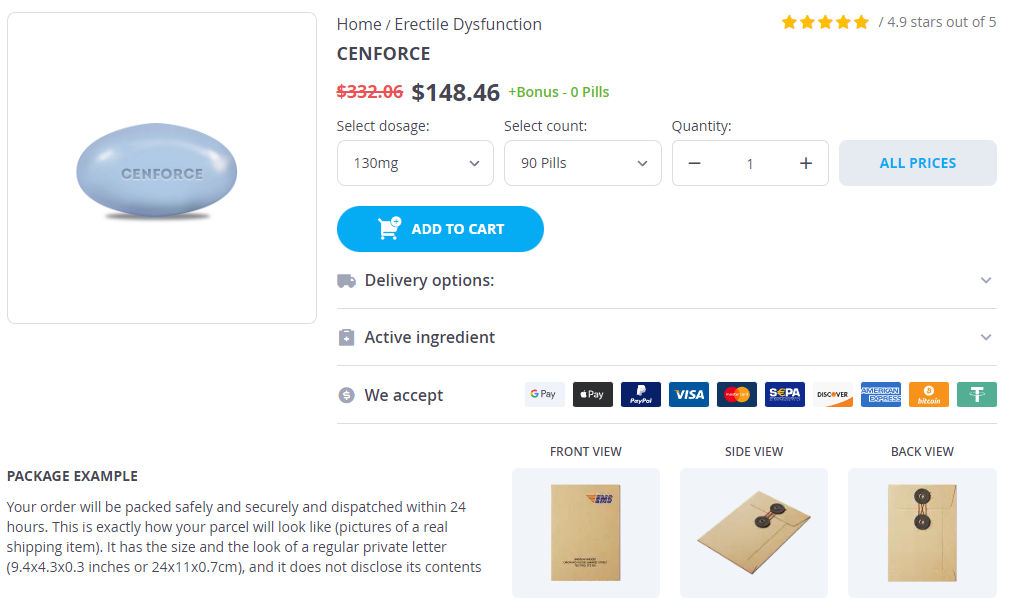
Cenforce dosages: 200 mg, 150 mg, 130 mg, 100 mg, 50 mg, 25 mg
Cenforce packs: 10 pills, 20 pills, 30 pills, 60 pills, 90 pills, 120 pills, 180 pills, 270 pills, 360 pills
In stock: 677
Only $0.56 per item
Description
When traced backwards this peritoneum is reflected on to the front of uterus at the junction of the body with the cervix treatment action group generic cenforce 130mg fast delivery. The posterior part of the superior surface of the bladder is in direct contact with the upper part of the cervix. The relations of the inferolateral surfaces of the bladder are the same as in the male except that the puboprostatic ligaments are replaced by the pubovesical ligaments. Ligaments of the Urinary Bladder the urinary bladder is kept in place by a number of so-called ligaments. The median umbilical ligament connects the apex of the urinary bladder to the umbilicus. It is the remnant of an embryonic structure the urachus that is derived from the allantoic diverticulum. The fascia over the upper surface of the levator ani (pelvic fascia) is thickened anteriorly to form the medial and lateral puboprostatic ligaments (in the male) or the pubovesical ligaments (in the female). Laterally the same fascia stretches from the bladder to the fascia covering the obturator internus. The lateral margins of the base of the bladder are joined to the lateral pelvic wall by fascia surrounding the veins that pass from the bladder to the internal iliac veins. The median umbilical ligament raises up a median fold of peritoneum called the median umbilical fold (33. In the fetus the right and left umbilical arteries pass from the internal iliac arteries to the umbilicus (on their way to the placenta). Their distal parts become obliterated and form the medial umbilical ligaments that connect the superior vesical arteries to the umbilicus. They raise up folds of peritoneum called the right and left medial umbilical folds. Peritoneum reflected from the superior surface of the bladder to the lateral wall of the pelvis is referred to as the lateral false ligament of the bladder. Two folds of peritoneum (right and left) pass backwards from the lateral margin of the base of the bladder to the sacrum. These folds pass lateral to the rectum and form the lateral boundaries of the rectovesical pouch. These folds are called the sacrogenital folds or the posterior ligaments of the bladder (33. On the posterior wall of the bladder, however, there is a triangular area where the mucosa is relatively fixed. The ureters open into the urinary bladder at the upper lateral corners of the trigone while the upper end of the urethra opens at the lower angle. The upper margin of the trigone forms a ridge stretching between the openings of the two ureters. The urinary bladder is supplied (in the male) by the superior and inferior vesical arteries. In the female the inferior vesical artery is replaced by the vaginal artery and the uterine artery also gives branches to the bladder.
Cinnamoni Cortex (Cassia Cinnamon). Cenforce.
- Loss of appetite, muscle and stomach spasms, bloating, intestinal gas, vomiting, diarrhea, common cold, impotence, bed wetting, menstrual complaints, chest pain, high blood pressure, kidney problems, cancer, and other conditions.
- Are there any interactions with medications?
- Diabetes.
- How does Cassia Cinnamon work?
- Dosing considerations for Cassia Cinnamon.
- Are there safety concerns?
- What is Cassia Cinnamon?
Source: http://www.rxlist.com/script/main/art.asp?articlekey=96963
The right border of the heart merges medicine 319 order 50mg cenforce with visa, above and below, with the corresponding vena cava. However, just lateral to the cardiac shadow irregular shadows are produced by structures in the hilum of each lung. That is why the structures belonging to the left half of the thorax are seen in the right half of the picture. An enlarged left atrium (abnormal) produces an indentation on the shadow of the oesophagus 470 Part 3 Thorax 23. A catheter was passed into the trachea, and then into the left principal bronchus, and a contrast medium was injected to outline the bronchi. The inner surfaces of the hip-bones are closely related to structures in the abdomen and pelvis. The lower ribs and costal cartilages give attachment to , and are related to , many structures in the abdomen. The cervical, thoracic and lumbar vertebrae can be easily distinguished from one another because of the following characteristics: 1. The transverse process of a cervical vertebra is pierced by a foramen called the foramen transversarium. These are present on the sides of the vertebral bodies and on the transverse processes. The pedicles are thick and short in the lumbar region and are directed backwards and somewhat laterally (24. The laminae of lumbar vertebrae are short and broad, but do not overlap each other. The inferior facets are slightly convex, and are directed equally forwards and laterally (24. Each superior articular process of a lumbar vertebra bears a rough projection called the mamillary process, on its posterior border. If the gap between the neural arches is small, no obvious deformity may be apparent on the surface (spina bifida occulta: occult = hidden). When the gap is large, meninges and nerves may bulge out through the gap forming a visible swelling. When neural elements are also present in the swelling the condition is called meningomyelocoele. Thefifthlumbarvertebramay be partially, or completely fused to the sacrum (sacralisation of 5th lumbar vertebra). However, when the abnormality described above is present, body weight can cause the body of the 5th lumbar vertebra to slip forward over the sacrum. Sometimes, a similar condition may affect the 4th lumbar vertebra that may then slip forwards over the 5th lumbar vertebra. Like other vertebrae those in the lumbar region can be fractured by direct injury.
Specifications/Details
The anterior ends of the ribs can move up or down by rotation at the costovertebral and costotransverse joints symptoms qt prolongation generic cenforce 130mg with mastercard. In expiration, the anterior ends of the ribs are lower than their posterior ends (17. During inspiration, the anterior end moves upwards in an arc becoming more horizontal. The forward movement of the rib is made possible by an angular movement at the manubriosternal joint. Rotation of ribs on a transverse axis takes place mainly in relation to the upper six ribs. These movements are facilitated by the fact that articular surfaces on the tubercles of these ribs are convex. The second movement of the ribs occurs on an axis that is roughly anteroposterior. During quiet breathing the movements of the ribs described above are produced by intercostal muscles. Elevation of ribs (during inspiration) is produced by the external intercostals, and depression (during expiration) by the internal intercostals, aided by elastic recoil of the thoracic wall. In deep inspiration movements of the ribs are aided by contraction of some muscles attached to the ribs. The scaleni (present in the neck) and the sternocleidomastoid muscles elevate the first rib, while the erector spinae helps expansion of the thorax by reducing the concavity of the thoracic part of the vertebral column. In forced inspiration (against resistance), the scapulae are elevated and fixed by the trapezius, the levator scapulae and the rhomboideus muscles. With the arms fixed (by holding onto a firm object) contraction of the serratus anterior and of the pectoralis major pulls upon the ribs helping expansion of the thorax. In forced expiration (as in patients with asthma), the thorax is compressed by the latissimus dorsi (but the major role is played by abdominal muscles). In infants, the thorax is more nearly circular as a result of which respiration is mostly abdominal. In a condition called emphysema the lungs are dilated, and as a result the thorax can become rounded in section (barrel chest), making respiration much less effective. Deformities seen in the thoracic cage may be congenital or may result from disease. In funnel chest, the front of the chest (in the region of the body of sternum and xiphoid process) is depressed. In pigeon chest, the thorax may project forwards in midline (as is normal in birds). In this chapter, we will consider other structures to be encountered in the thoracic wall.
Syndromes
- Heart failure
- Adults who wear contact lenses need yearly eye exams
- Blindness or severe visual disability
- Delay in clamping the umbilical cord
- Tremors
- Urinary tract infection
- Stress incontinence
- Failure of the lung to expand
- What medications do you take?
Related Products
Additional information:
Usage: gtt.
Tags: generic cenforce 130mg buy, quality 25 mg cenforce, order 130mg cenforce amex, generic 200mg cenforce overnight delivery
9 of 10
Votes: 293 votes
Total customer reviews: 293
Customer Reviews
Basir, 33 years: In some cases chronic respiratory obstruction leads to considerable dilatation of alveoli. It ends by supplying the skin of the upper and medial part of the thigh, over the pubis and the adjoining part of the genitalia. This is easier if a segment of the ductus deferens has not been removed during vasectomy. Multiple image views should confirm the guide pin position be- fore inserting the bone screw to avoid stress risers from too many holes in the bone.
Harek, 39 years: The Anal Musculature the anal canal is surrounded by a number of sphincters that are as follows (33. Before proceeding to study the features to be seen on individual surfaces of the liver, it is necessary to briefly consider the basic peritoneal relationships of the organ. The sheaths for the tendons going to the digits, and that for the extensor pollicis longus, extend to the level of the middle of the metacarpus. Plantar flexion of distal Tibial nerve (S2, 3) medial malleolus and phalanges then on medial side of 2.
Yasmin, 60 years: The loose (or flail) segment moves inwards with inspiration and outwards during expiration. Abnormal development of gonads and genitalia gives rise to various types of hermaphroditism, in which there is a mixture of male and female characters. In the female the corresponding fibres pass across the sides of the vagina to end in the perineal body. There is marked asymmetry of the mouth because of paralysis of the orbicularis oris and of muscles inserted into the angle of the mouth.
Lars, 36 years: In maxillary sinusitis there may be tenderness over the cheek below the inferior orbital margin. Crossing the median plane carry the line to the right costal margin which it should cut at the level of the tip of the ninth costal cartilage. We have seen that normally the pleural cavity is a potential space containing a thin film of serous fluid that separates visceral and parietal pleura. The porta hepatis and the fissure for the ligamentum venosum give attachment to the two layers of the lesser omentum.
Silvio, 51 years: Infection in any part of the upper limb can lead to lymphadenitis or lymphangitis. Lee A, Driscoll D, Gloviczki P, Clay R, Shaughnessy W, Stans A: Evaluation and management of pain in patients with Klippel-Trenaunay syndrome: a review. This ganglion is located in the pterygopalatine fossa and is suspended from the maxillary nerve by two ganglionic branches (43. The lesion is more effectively excised by a lateral rhinotomy or midface degloving approach.
-
Our Address
-
For Appointment
Mob.: +91-9810648331
Mob.: +91-9810647331
Landline: 011 45047331
Landline: 011 45647331
info@clinicviva.in -
Opening Hours
-
Get Direction


