
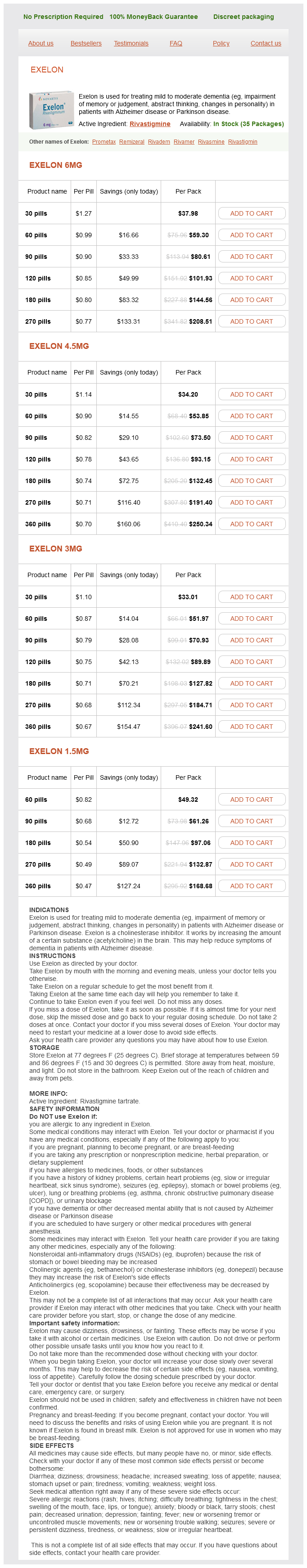
Exelon dosages: 6 mg, 4.5 mg, 3 mg, 1.5 mg
Exelon packs: 30 pills, 60 pills, 90 pills, 120 pills, 180 pills, 270 pills, 360 pills
In stock: 818
Only $0.5 per item
Concurrent inflammation of both eyes occurs in the majority of patients with frequent foveal involvement medications quizlet discount exelon 6 mg buy on line. However, the lesions demonstrate growth in size and contiguous relapses over several months. Lesions are hypofluorescent in the early phase due to blockage or choriocapillaris nonperfusion. Later, hyperfluorescence develops, starting at the borders of the lesion, with uniform late staining of the underlying fibrotic scar and sclera. The authors concluded that an antibiotic could possibly act as an antigenic trigger for this disease. Over the course of several weeks, the acute lesions first grow in size and then evolve to inactive pigmented chorioretinal scars. Corticosteroids have been used with rapid resolution of active lesions and improvement in vision. Yet, it is difficult to conclude that steroids affected the long-term outcome, given the natural history of spontaneous healing of individual lesions. Antiviral agents also have been tried in a small number of patients but without obvious benefit to date. Other immunosuppressive agents, such as cyclosporine, have also been used,115 but recurrence often is seen after immunosuppression is tapered. They are usually clinically distinguishable based on specific characteristics including age of onset, course, visual outcome, systemic associations, and recurrence patterns. Given the clinical and angiographic similarities among these entities, they may share a common immune dysregulation. The present work was supported in part by an unrestricted grant from Research to Prevent Blindness, Inc, New York City. Bonnin P, Lavat P: In connection with a case of Gass placoid epitheliopathy in 1860. Bohlender T, Weindler J, Ratzkova A: Indocyanine green angiography in acute posterior multifocal placoid pigment epitheliopathy. Schneider U, Inhoffen W, Gelisken F: Indocyanine green angiography in a case of unilateral recurrent posterior acute multifocal placoid pigment epitheliopathy. Yuzawa M, Kawamura A, Matsui M: Indocyanine green videoangiographic findings in acute posterior multifocal placoid pigment epitheliopathy. Isashiki M, Koide H, Yamashita T, et al: Acute posterior multifocal placoid pigment epitheliopathy associated with diffuse retinal vasculitis and late haemorrhagic macular detachment. Sigelman J, Behrens M, Hilal S: Acute posterior multifocal placoid pigment epitheliopathy associated with cerebral vasculitis and homonymous hemianopia. Bewermeyer H, Nelles G, Huber M, et al: Pontine infarction in acute posterior multifocal placoid pigment epitheliopathy. Fujisawa C, Fujiwara H, Hasegawa E, et al: the cases of serpiginous choroiditis [Japanese, English abstract].
Myrica pensylvanica (Bayberry). Exelon.
Source: http://www.rxlist.com/script/main/art.asp?articlekey=96199
Tonography may occasionally play a role in the diagnosis and management of chronic angle-closure glaucoma treatment xanthelasma eyelid exelon 4.5 mg purchase overnight delivery. With compression, the angle opens and peripheral anterior synechiae are identified. Displacement of focal lines indicates that an open space exists between the iris periphery and the chamber angle wall. Focal lines are not displaced but form an angle, the apex corresponding to the point of contact between the anterior and the posterior walls of the chamber angle. Low, diffuse synechial closure, typical of primary chronic angle-closure glaucoma. Primary chronic angle-closure glaucoma is most frequently mistaken for primary open-angle glaucoma. The previous discussion has emphasized the importance of gonioscopy in distinguishing between these two entities. If the posterior trabeculum is observed, the examiner should suspect that a narrow angle is contributing to the decrease in outflow facility. If synechiae are noted and are not secondary to another disease or previous surgical procedure, primary chronic angleclosure exists. In blue-eyed patients, this meshwork lines the angle overlying the ciliary body band, scleral spur, and trabecular meshwork. It appears as a delicate gray or colorless structure that interferes with the visibility of the angle. Typically, the anterior edge of the uveal meshwork is on the trabecular meshwork and has many fine branching twiglike endings. The feature that seems to be the most consistent in distinguishing normal tissue from peripheral anterior synechiae is a difference in the apparent solidity of tissue. The uveal meshwork in adult eyes regularly has the lacy, open character of a meshwork made up of many interconnected strands. Peripheral anterior synechiae may occur in other conditions and may mimic primary chronic angle-closure glaucoma. It must also be remembered that any condition that secondarily causes shallowing of the anterior chamber may cause permanent synechial closure. These conditions include both inflammatory and postsurgical conditions in which adhesions may develop between the iris, vitreous face, or intraocular lens. Pupillary block occurs most often after a complicated cataract extraction but may also be noted in a quiet eye with an intact posterior capsule and a well-positioned posterior chamber intraocular lens. Unfortunately, we are all too familiar with shallow anterior chambers secondary to filtering surgery. Peripheral anterior synechiae that occur after prolonged absence of the anterior chamber after surgery are usually broad bands of adhesions to the iris at various levels.
Vendantham V: Optical coherence tomography findings in macular hole due to argon laser burn treatment for bronchitis buy 1.5 mg exelon fast delivery. Niwa H, Tersasaki H, Ito Y, Mikake Y: Macular hole development in fellow eyes with unilateral macular hole. Kuhnt H: Ueber eine eigenthumliche Veranderung der Netzhaut as maculam (retinitis atrophicans sive rareficans centralis). GlacetBernard A, Zourdani A, Perrenoud F, et al: Stage 3 macular hole: role of optical coherence tomography and B-scan ultrasonography. Mori K, Abe T, Yoneya S: Dome shaped detachment pf premacular vitreous cortex in macular hole development. Altaweel M, Ip M: Macular hole: improved understanding of pathogenesis, staging, and management based on optical coherence tomography. Twoyear results of a randomized clinical trial comparing natural history, vitrectomy, and vitrectomy plus autologous serum: Moorefields Macular Hole Study Group Report No. Paques M, Chastang C, Mathis A, et al: Effect of autologous platelet concentrate in surgery for idiopathic macular hole: results of a multicenter, double-masked, randomized trial. Hoerauf H, Kluter H, Joachimmeyer E, et al: Results of vitrectomy and the notouch-technique using autologous adjuvants in macular hole treatment. Mester V, Kuhn F: Internal limiting membrane removal in the management of full-thickness macular holes. Gandorfer A, Haritoglou C, Kampic A: Retinal damage from indocyanine green in experimental macular surgery. Li K, Wong D, Hiscott P, et al: Trypan blue staining of internal limiting membrane and epiretinal membrane during vitrectomy: visual results and histopathological findings. Horio N, Horiguchi M, Yamamoto N: Triamcinolone-assisted internal limiting membrane peeling during idiopathic macular hole surgery. Krohn J: Duration of face-down positioning after macular hole surgery; a comparison between 1 week and 3 days. Hirata A, Yonemura N, Hasamura T, et al: Effect of infusion pressure on visual field defects after macular hole surgery. Choroidal folds develop secondary to biomechanical stresses, for instance, from an extraocular mass in the orbit pressing upon the globe, from thickening of the choroid from hypotony, choroidal effusion, or inflammation, or from stresses secondary to a growing choroidal tumor. Both choroidal and retinal folds can be seen upon biomicroscopy but fluorescein angiography outlines choroidal folds most dramatically. The clinical presence of choroidal or retinal folds needs to be explained, as the underlying disease which created them may need to be addressed. Both types of folds can be symptomatic with retinal folds creating metamorphopsia. This article details the biomechanical principles which create chorioretinal folds so they can be better understood. Many disorders are associated with the formation of folds in the choroid and retina. Choroidal folds are often a sign of orbital or ocular disease, but they may develop after surgery on the eye or orbit or may occur idiopathically.
Syndromes
Additional information:
Usage: p.c.
Tags: discount exelon 4.5 mg on line, cheap exelon 3 mg mastercard, 3 mg exelon mastercard, buy exelon 1.5 mg low cost
Stejnar, 52 years: One other point that bears mentioning is the reported central nervous system effects of topical b-blockers, such as depression, lethargy, confusion, and hallucinations.
Nasib, 63 years: A Barkan goniotomy lens modified with an added handle, and placed onto a mound of healon on the central cornea works well.
Jensgar, 36 years: Four of 10 patients eventually experienced subretinal neovascularization in association with perifoveal scars.
Kaffu, 54 years: The widely quoted study of Folk et al15 retrospectively studied 388 consecutive cases in which phakic retinal detachment associated with lattice degeneration occurred in one eye and lattice degeneration was present in the second eye.
Gunock, 31 years: Takahashi T et al: A clinical evaluation of uveitis-associated secondary glaucoma.
C-585, Saraswati Vihar, Pitampura, New Delhi 110034


