
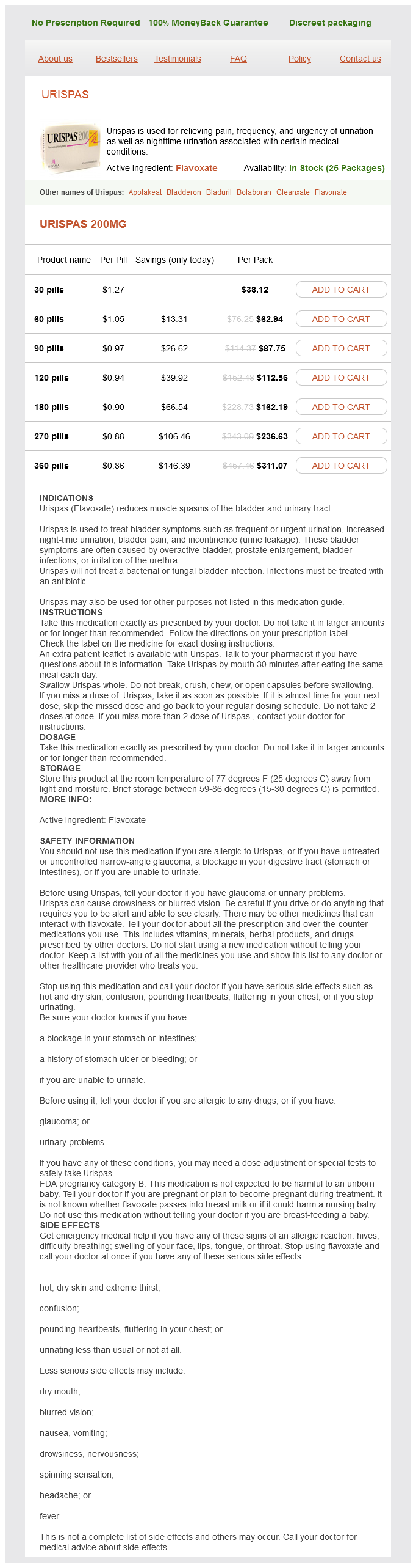
Flavoxate dosages: 200 mg
Flavoxate packs: 30 pills, 60 pills, 90 pills, 120 pills, 180 pills, 270 pills, 360 pills
In stock: 996
Only $0.92 per item
Small resting lymphocytes spasm effective 200 mg flavoxate, stromal elements, and plasma cells fill the interfollicular zone, which may also contain patchy accumulations of histiocytes. The immunoglobulin Dpositive mantle should be present and fairly crisply demarcated from the immunoglobulin Dnegative germinal center inside and the immunoglobulin Dnegative paracortex beyond. Because of the close overlap with marginal zone lymphoma, even when the immunoarchitecture is reassuringly intact, flow cytometry or molecular studies are needed to confirm the polyclonal nature of the process. Marginal zone lymphoma is the most challenging element of the differential diagnosis. Some patients have systemic symptoms, such as fever and weight loss, and many (70%) have polyclonal hypergammaglobulinemia; both the clinical and laboratory abnormalities likely reflect the underlying altered immune state. Small, cytologically bland lymphocytes and intermingled plasma cells distend the alveolar walls, with accentuation along the bronchovascular bundles and lobular septae (Box 16. Aggregates of histiocytes or poorly formed granulomas may be present, but neutrophils and eosinophils are scarce. Hypersensitivity pneumonitis may lead to cellular interstitial infiltrates, but on scanning view, the process should show bronchocentricity, and loosely formed granulomas or at least multinucleated giant cells are likely to be more prominent. Some patients may have spontaneous resolution, and others achieve a good response to a trial of steroids. Morbidity and mortality are most often seen in patients with superimposed infection or other comorbid conditions, such as renal failure. One, the hyaline vascular variant, almost invariably presents via mass effect of a solitary mediastinal mass or central lymphadenopathy in otherwise asymptomatic individuals and is cured by complete resection. The other, the plasma cell variant, has protean manifestations and multifocal disease and has significant associated morbidity and mortality. What brings them together85 is that, in historical examples, the two histologies may coexist in the same lymph node, and second, they were characterized in separate publications by the same individual, Benjamin Castleman. At low power, the architecture is nodular, composed of small and involuted germinal centers with expansive immunoglobulin Dpositive mantles formed of small lymphocytes, often in a laminated or "onion skinning" array. Multiple germinal centers may be found in the boundaries of a single mantle zone, and in opportune sections, the lymphoid depletion in the germinal centers unmasks the radially penetrating high endothelium, the "lollipop" motif (Box 16. Sinuses between the regressed follicles are imperceptible, usually because they are absent. At high power, the germinal centers are depleted of lymphocytes and contain both extracellular matrix and abundant follicle dendritic cells. The interfollicular zone includes plasmacytoid monocytes, stromal myoid cells, histiocytes, dendritic cells, and lymphocytes.
Calendula. Flavoxate.
Source: http://www.rxlist.com/script/main/art.asp?articlekey=96263
Posterior fossa tumor with distinct choroid plexus papilloma and ependymoma components muscle relaxer sleep aid flavoxate 200 mg buy with amex. Prognostic implications of atypical histologic features in choroid plexus papilloma. The cytologic findings in choroid plexus carcinoma: report of a case with differential diagnosis. Villous hypertrophy versus choroid plexus papilloma: a case report demonstrating a diagnostic role for the proliferation index. A potentially useful aid in differentiating carcinoma of the choroid plexus from metastatic papillary carcinomas. Usefulness of synaptophysin immunohistochemistry in an adult case of choroid plexus carcinoma. Choroid plexus tumors differ from metastatic carcinomas by expression of the excitatory amino acid transporter-1. Choroid plexus tumors in children: immunohistochemical and scanning electron-microscopic features. Comparative genomic hybridization detects specific cytogenetic abnormalities in pediatric ependymomas and choroid plexus papillomas. High-resolution genomic analysis does not qualify atypical plexus papilloma as a separate entity among choroid plexus tumors. Choroid plexus carcinomas are characterized by complex chromosomal alterations related to patient age and prognosis. Platelet-derived growth factor receptor expression and amplification in choroid plexus carcinomas. Methylation profiling of choroid plexus tumors reveals 3 clinically distinct subgroups. Use of ifosfamide, carboplatin, and etoposide chemotherapy in choroid plexus carcinoma. Radiation therapy for choroid plexus carcinoma patients with Li-Fraumeni syndrome: advantageous or detrimental Nearly all patients have a long-standing history of epilepsy that is refractory to medical treatment. Introduction Not all brain tumors fit neatly into well-defined diagnostic categories within accepted classification systems. Such is the case with the three rare neoplasms discussed in this chapter: angiocentric glioma, astroblastoma, and chordoid glioma. Each of these has a unique and readily recognized histologic pattern, variably defining molecular and immunohistochemical features, and occurs within a specific clinicopathologic setting. These features also set them apart from the more frequent astrocytic and ependymal tumors with which they are often confused. Radiologic Features and Gross Pathology Tumors are typically centered in the cortex, but often extend into subcortical regions of the frontal, temporal, or parietal lobes.
Acute disseminated encephalomyelitis in the intensive care unit: clinical features and outcome of 20 adults spasms 5 month old baby order 200 mg flavoxate with visa. Cultivation of papova-like virus from human brain with progressive multifocal leukoencephalopathy. Clinical outcomes of natalizumab-associated progressive multifocal leukoencephalopathy. Central nervous system immune reconstitution disease in acquired immunodeficiency syndrome patients receiving highly active antiretroviral treatment. A matrix-less measles virus is infectious and elicits extensive cell fusion: consequences for propagation in the brain. Vacuolar myelopathy pathologically resembling subacute combined degeneration in patients with the acquired immunodeficiency syndrome. Leukodystrophies: pathogenesis, diagnosis, strategies, therapies, and future research directions. Peroxisomal disorders: genotype, phenotype, major neuropathologic lesions, and pathogenesis. Diffuse progressive leukodystrophy in the adult with production of metachromatic degenerative products (Alzheimer-Baroncini). Significance of sulfatide and other lipid abnormalities in white matter and kidney. Progressive fibrinoid degeneration of fibrillary astrocytes associated with mental retardation in a hydrocephalic infant. Glia-specific activation of all pathways of the unfolded protein response in vanishing white matter disease. Foamy cells with oligodendroglial phenotype in childhood ataxia with diffuse central nervous system hypomyelination syndrome. Central pontine myelinolysis: a hitherto undescribed disease occurring in alcoholic and malnourished patients. Processing Epilepsy Surgery Specimens Epilepsy-related resections are among some of the more challenging surgical neuropathology specimens to evaluate. A systematic and careful approach to dissecting and evaluating these cases is important in optimizing diagnostic capabilities. An attempt should be made to orient the specimen with respect to location-posterior versus anterior, medial versus lateral. Close inspection of the exterior appearance of the excised tissue should be performed. Abnormalities such as Sturge Weber, gyral abnormalities, certain superficially situated tumors such as dysembryoplastic neuroepithelial tumors, and contusion-related damage related to invasive seizure monitoring may be evident. The specimen should be carefully sectioned at approximately 5-mm parallel intervals according to the orientation, ideally in coronal plane along the anterior-posterior axis. The cross sections should be carefully inspected for abnormalities, which should be documented. Look for blurring of the gray-white interface, discoloration, cystic lesions, and abnormal architecture.
Syndromes
Additional information:
Usage: gtt.
Tags: cheap 200 mg flavoxate fast delivery, flavoxate 200 mg order visa, 200 mg flavoxate purchase with amex, flavoxate 200 mg without prescription
Yugul, 33 years: As previously discussed, crushed samples of carcinoid and especially atypical carcinoid should be considered, and some advocate Ki-67 in all cases in which morphology is limited. Rathke cysts in the sellar region, colloid cysts in the third ventricle, and neurenteric cyst in the ventral cervical spinal cord show similar histologic features of pseudostratified, ciliated, columnar epithelium with goblet cells. Discussions with neurologists and neuroradiologists and examination of serial sections of paraffin blocks, as well as analysis of molecular pathology for monoclonal B and T cell rearrangement are required in these cases (see Chapter 26). Practical Pulmonary Pathology Clinical Presentation Sarcoidosis is a systemic disease of uncertain etiology and with frequent lung manifestations.
Gembak, 34 years: There are foci or areas of conspicuous keratinization of tumor cells admixed within the nests of polygonal cells as described above. Ancillary Diagnostic Studies If possible, frozen section of either fresh or formalin-fixed tissue should be performed and stained with a neutral fat-soluble dye such as Oil Red O, which will demonstrate the intravascular fat emboli. Diagnosis Hyaline membrane disease with acute pulmonary interstitial emphysema Discussion Hyaline membrane disease is the pathologic correlate of respiratory distress syndrome in the neonatal period. Entrapped intratumoral axons are few, often subcapsular, and seen only on special stains.
Topork, 52 years: The previously published view that brain invasion in meningiomas represents the most reliable sign of malignancy is no longer tenable. Which of the following is/are histopathologic feature(s) in lung biopsies that favor infection over acute rejection Neuropathological findings in primary generalized epilepsy; a study of eight cases. Before any wedge lung biopsy is performed, consultation among the radiologist, chest physician, and thoracic surgeon is essential to ensure appropriate sampling and the identification of ideal locations for biopsy.
Dargoth, 43 years: Findings can include fever, headache, obtundation, hemiparesis, seizures, or other focal deficits. Cytomegalovirus pneumonia in bone marrow transplant recipients: miliary and diffuse patterns. Time Period Historically, infections have been most common around the seventh posttransplantation week. The nodules may be present anywhere within the lung parenchyma but typically are most numerous in the upper lung zones.
Bogir, 23 years: Prognosis is generally poor, with development of chronic lung disease or death in a majority of affected infants. A practical approach to biopsies with dense cellular infiltrates is provided at the end of this chapter. In the United States, local transmission has been documented in Florida, Puerto Rico, and the U. Lipomas with complex elements (lipomatous hamartomas) should be distinguished from teratomas, the latter containing a more complex mixture of all three germinal layers.
Masil, 47 years: Intra-abdominal pulmonary sequestration associated with congenital cystic adenomatoid malformation of the lung: just an unusual combination of rare pathologies Pulmonary alveolar proteinosis: clinical aspects and current concepts in pathogenesis. Pulmonary toxicity has been reported in 5% to 10% of patients taking this medication. This set of findings reflects the fact that plexiform lesions, in our view, are actually the result of necrosis of the vessel, with subsequent thrombosis and organization.
C-585, Saraswati Vihar, Pitampura, New Delhi 110034


