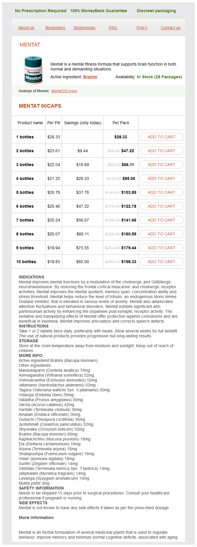
Mentat
Mentat
Mentat dosages: 60 caps
Mentat packs: 1 bottles, 2 bottles, 3 bottles, 4 bottles, 5 bottles, 6 bottles, 7 bottles, 8 bottles, 9 bottles, 10 bottles
In stock: 673
Only $21.07 per item
Description
Some stroke centers have a standardized cutoff for candidates undergoing thrombectomy everlast my medicine cheap mentat 60 caps mastercard, where candidates with poor scores (often less than 6, with no ischemic changes scoring 10) are being excluded from thrombectomy because of the documented poor outcomes in this group of patients, despite successful revascularization. Likewise, a significant lack of collateralization on delayed arterial imaging may indicate a poor outcome in this patient population despite being within the time window. Further studies are ongoing and, if shown to be beneficial, may ultimately become the standard of care in determining candidacy for thrombectomy. These are qualitative in their assessment of cerebral blood flow after processing contrast arrival and washout times. This leads to a wide variation in the data, and interpretation is therefore useful only for symmetry assessment. Current guidelines do not require perfusion information to guide therapy, but will likely also play a role in the future. Based on the stroke guidelines, permissive hypertension is the standard of care, but care must be taken to avoid hypotension, as this could lead to further secondary ischemic injury to the penumbra tissue. Osmotic therapies have limited utility in ischemic infarcts, as the dead tissue does not respond to diuresis, whereas normal brain parenchyma does, which can theoretically worsen shift and herniation. Current standards aim at maintaining sodium levels in the high normal range, thus avoiding hyponatremia. Normothermia is recommended to minimize secondary injury, but there is no evidence to suggest that therapeutic cooling in ischemic stroke improves overall outcome. Cerebral metabolism increases with fever, requiring more blood flow to the already hypoperfused tissue, thus hyperthermia must be avoided to minimize secondary ischemic injury. Patients who develop large hemispheric infarctions despite best medical interventions may progress to develop significant edema, as the dead tissue accumulates cytotoxic fluid. In the elderly population, this can often be tolerated because of prior diffuse cerebral volume loss. However, in younger patients, edema can lead to severe hemispheric swelling with transtentorial or uncal herniation, followed by brainstem compression, coma, and death. Evidence suggests that early decompression with hemicraniectomy with or without removing some of the necrotic tissue leads to improved outcomes. Often surgeons will remove the anterior temporal lobe to reduce lateral brainstem compression. If clinical deterioration occurs, there is an increase in morbidity and mortality. Recent randomized controlled trials performed across Europe demonstrated markedly improved survival after hemicraniectomy in this population [8]. Patients older than 60 years have had even worse outcomes in one randomized controlled trial, with 93% of the surviving patients requiring significant or complete care with daily living needs [9]. Patients with large cerebellar ischemic insults with little brainstem involvement are good surgical candidates for decompression and removal of ischemic tissue to prevent imminent direct brainstem compression and the potential for rapid deterioration. Facial and sixth nerve palsies can be early signs of significant brainstem compression that may rapidly be followed by a loss of brainstem function. Placement of an external ventricular drain to treat acute hydrocephalus combined with posterior fossa craniectomy often results in rapid return to baseline neurological function.
Neuromins (Dha (Docosahexaenoic Acid)). Mentat.
- How does Dha (docosahexaenoic Acid) work?
- What other names is Dha (docosahexaenoic Acid) known by?
- Attention deficit-hyperactivity disorder (ADHD).
- Depression, dementia, improving vision, high cholesterol, improving infant development, reducing aggressive behavior in people under stressful situations, improving night vision in children with dyslexia, improving movement disorders in children, and other conditions.
- Depression.
- Type 2 diabetes.
- Reducing the risk of death in people with coronary artery disease, when DHA is consumed as part of the diet.
- Psoriasis.
Source: http://www.rxlist.com/script/main/art.asp?articlekey=96835
Imaging provides detailed information on brain tissue and vascular anatomy for clinicians to corroborate their clinical history and examination findings in acute stroke cases medications jaundice cheap mentat 60 caps with mastercard. Transesophageal-echocardiography-facilitated early cardioversion from atrial fibrillation: short-term safety and impact on maintenance of sinus rhythm at 1 year. Prevalence of residual left atrial thrombi in patients presenting with acute thromboembolism and newly recognized atrial fibrillation. The veterans affairs stroke prevention in nonrheumatic atrial fibrillation investigators. The significance of imaging in stroke depends on unraveling the pathophysiology of acute ischemia and plays a pivotal role during the acute phase to formalize therapeutic plans. Across a variety of practice scenarios, clinicians often have a choice of various imaging modalities to yield further information on the care of patients with stroke. Imaging data serve as an extension of the clinical examination that routinely enhances the clinical evaluation and neurological localization in a patient with stroke. The following discussion delineates the key goals of neuroimaging in stroke to augment clinical decision making. It has been shown that time alone is rudimentary and neuroimaging complements decision making in acute stroke cases. These imaging strategies have been used on a regular basis as they portray a snapshot of brain parenchyma and guide further therapeutic decisions. Imaging in stroke focuses to determine the diagnosis and etiology, lesion localization, extent of ischemic evolution, therapeutic implications, and expected prognosis. Rather than focusing solely on ruling out massive infarcts, the trend has shifted to evaluate subtle signs of ischemia including hypoattenuation or obscuration of the lentiform nuclei, loss of the insular ribbon, sulcal effacement, cortical hypodensity, and the presence of various hyperdense vessel findings. Neuroimaging usually compliments the examination findings obtained by clinicians, whereas lesion patterns are particularly helpful in stroke. The proximal nidus or embolic source may be arteryartery or from the heart, prompting further diagnostic testing. Border zone lesions of ischemia between the principal arterial territories in the brain may manifest as a string or archipelago of discrete cortical and subcortical lesions that implicate a proximal stenotic or occlusive lesion causing hypoperfusion. Contrast enhancement of ischemic lesions is helpful in the age determination of any vascular lesion. Parenchymal enhancement is associated with disruption of the bloodbrain barrier that usually presents in a gyriform or ring-like shape, located peripheral to the central ischemic lesion. It usually becomes visible around 47 days after the ischemic event, tends to resolve within 8 weeks, and may persist beyond 3 months in isolated cases. Serial imaging of infarct evolution over ensuing days or weeks illustrates the dynamic nature involved in cerebral ischemia and various stages may be encountered, including "fogging" or the transient disappearance of subacute lesions before chronic scar formation. Atherosclerotic plaques are the most important culprits leading to stenosis or distal thromboembolic phenomenon due to plaque rupture. Carotid ultrasound has been used for decades to assess intimal thickness and calculate focal stenosis in extracranial circulation.
Specifications/Details
Medical therapies without the goal of reperfusion for massive infarction of the cerebral hemispheres or the cerebellum are directed toward preventing additional ischemic injuries and controlling brain edema medicine quetiapine 60 caps mentat with visa. Depending on the cause of vascular occlusion, anticoagulation is often indicated to prevent additional embolic events or thrombus propagation. A moderately elevated perfusion pressure must be maintained while avoiding both hypotension and excessive hypertension. Ideally, at least in the initial days, this should take place in a neurointensive care unit. Traditionally, osmotic diuretics administration and intubation combined with hyperventilation have been used when the mass effect is accompanied by a decreased mental status. Despite such therapeutic efforts, 2030% of massive infarctions resulted in irreversible secondary brainstem injuries caused by herniation and mass effect [5]. In some cases, and to prevent these secondary brainstem injuries, decompressive surgery is suitable. On the other hand, if bone flap replacement is delayed further, communicating hydrocephalus may develop requiring shunt placement [7]. The most commonly described signs of deterioration from hemispheric supratentorial infarction are ipsilateral pupillary dysfunction, varying degrees of mydriasis, and ocular adduction paralysis. A false-localizing Babinski sign as a result of brainstem compression against the contralateral tentorium (Kernohan notch) can also occur. Abnormal respiratory patterns reflecting brainstem dysfunction typically occur late in the course; these include central neurogenic hyperventilation or ataxic respiratory patterns and periodic breathing [9]. Of note, patients with significant preexisting morbidities were excluded from this analysis. In this pooled analysis, hemicraniectomy was associated with a statistically significant reduction of mortality, from 75% to 24%. Hemicraniectomy was found to be associated with a statistically significant reduction in mortality, from 60. As for younger patients, hemicraniectomy was associated with a significant reduction in mortality (from 76% to 43%). Severe depression was present in 80100% of the survivors, irrespective of intervention. This has led some providers to decide not to perform surgery in this patient population if the dominant hemisphere is affected. Also, additional reasonable interpretation of the available data sets is that surgery may reduce morbidity in select patient populations (younger than 60 years) when performed within 48 h. On the other hand, surgical treatment increased the probability of being fully independent from 2% to 14% and that of having no/minimal dependency from 21% to 43%. Close neurologic and cardiovascular monitoring in an intermediate or intensive care stroke unit in patients with territorial cerebellar infarctions for up to 5 days is recommended, even if the patient seems to be stable [13].
Syndromes
- If you are or might be pregnant
- Hypothyroidism
- Poor alignment of the toe
- Hormone replacement therapy and estrogens
- Pain with intercourse
- Certain measures, such as fruit juice or honey, have been recommended to treat a hangover. But there is very little scientific evidence to show that such measures help. Recovery from a hangover is usually just a matter of time. Most hangovers are gone within 24 hours.
- Drug abuse
- Anti-A serum, you have type A blood
- Weakness of the face
Related Products
Additional information:
Usage: p.o.
Tags: cheap mentat 60 caps fast delivery, buy mentat 60 caps, generic 60 caps mentat with amex, mentat 60 caps purchase line
9 of 10
Votes: 89 votes
Total customer reviews: 89
Customer Reviews
Pakwan, 51 years: Selected cerebral infections that have been associated with stroke and vasculitis are listed in Table 124.
Ford, 33 years: Those who present asymptomatically and do not progress to growth have a relatively benign course with 0.
Kliff, 24 years: Dystrophic microcalcifications are common in chronic established lesions and a peripheral rim of hemosiderin-storing macrophages is present in the adjacent gliotic brain.
Temmy, 47 years: Enzymes are present in the vessel that degrade substances and prevent them from crossing the capillary.
Kafa, 64 years: Cigarettes and Ischemic Stroke the Framingham study demonstrated that cigarette smoking increases risk of all types of stroke due to arterial thromboembolism, is dose related, and independent of history of hypertension [56,57].
-
Our Address
-
For Appointment
Mob.: +91-9810648331
Mob.: +91-9810647331
Landline: 011 45047331
Landline: 011 45647331
info@clinicviva.in -
Opening Hours
-
Get Direction


