
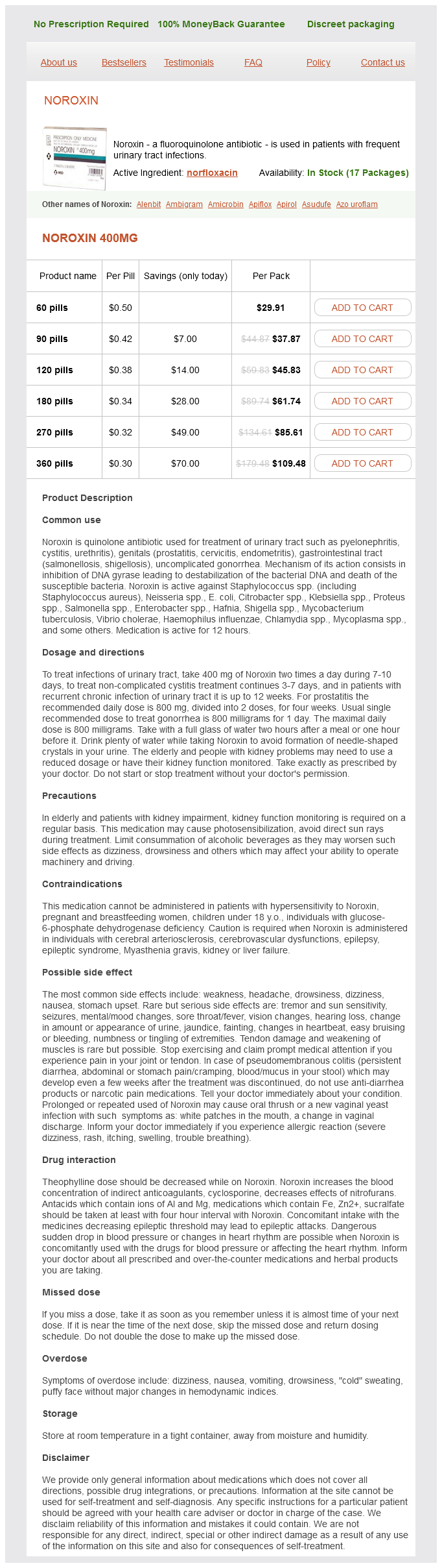
Norfloxacin dosages: 400 mg
Norfloxacin packs: 60 pills, 90 pills, 120 pills, 180 pills, 270 pills, 360 pills
In stock: 626
Only $0.32 per item
Langerhans cell histiocytosis antimicrobial vs antibiotics buy 400 mg norfloxacin otc, 456-459 - diagnostic checklist, 457 - differential diagnosis, 457 - eosinophilic pneumonia vs. Large cell neuroendocrine carcinoma, 155 - poorly differentiated small cell variant squamous carcinoma vs. Lipoid pneumonia (paraffinoma), 324-327 - diagnostic checklist, 325 - differential diagnosis, 325 - histochemical features, 325 Lipoma - bronchial, cartilaginous hamartoma vs. Lung neoplasms, malignant, primary - acinic cell carcinoma, 198-205 diagnostic checklist, 199 differential diagnosis, 199 granular cell tumor vs. Lymphoblastic lymphoma, 748-753 - diagnostic checklist, 750 - differential diagnosis, 750 - genetic testing, 749 - immunohistochemistry, 750 - intrapulmonary thymoma vs. Mediastinal foregut cyst, benign, 870-875 - diagnostic checklist, 871 - differential diagnosis, 871 - histochemical features, 872 - immunohistochemistry, 872 - prognosis, 871 Mediastinal germ cell tumors, clinical staging, 710, 716 Mediastinal Hodgkin lymphoma, 726-733 - diagnostic checklist, 728 - differential diagnosis, 727-728 - immunohistochemistry, 728 - mediastinal diffuse large cell lymphoma vs. Mesothelial hyperplasia, 554-557 - diagnostic checklist, 555 - differential diagnosis, 555 - prognosis, 555 Mesothelioma, malignant, 576-583 - biphasic pleural endometriosis vs. Metastasizing leiomyoma, benign, 352-355 - differential diagnosis, 353 - prognosis, 353 Metastatic calcification. Mucinous type, bronchioloalveolar carcinoma, 89 Mucoepidermoid carcinoma, 186-191 - adenoid cystic carcinoma vs. Mucous gland adenoma, 12-17 - diagnostic checklist, 14 - differential diagnosis, 13-14 - histochemical features, 14 - immunohistochemistry, 14 - molecular features, 14 - mucoepidermoid carcinoma vs. Myoepithelial carcinoma of lung, 212-215 - diagnostic checklist, 213 - differential diagnosis, 213 - prognosis, 213 Myofibroblastic tumor. Peripheral nerve sheath tumors, 790-795 - diagnostic checklist, 792 - differential diagnosis, 791-792 - immunohistochemistry, 792 - malignant malignant solitary fibrous tumor vs. Pleural neoplasms, benign - calcified fibrous pseudotumor, 568-571 diagnostic checklist, 569 differential diagnosis, 569 prognosis, 569 - mesothelial hyperplasia, 554-557 diagnostic checklist, 555 differential diagnosis, 555 prognosis, 555 - solitary fibrous tumor, 558-567 diagnostic checklist, 560 differential diagnosis, 559-560 genetic testing, 559 histologic growth patterns, 560 immunohistochemistry, 560 prognosis, 559 Pleural neoplasms, malignant, 574-575 - molecular genetic alterations, 575 - primary, 574 - prognostic factors, 575 - secondary, 574 - staging, 575 Pleural neoplasms, malignant, primary - angiosarcoma, 596-601 diagnostic checklist, 597 differential diagnosis, 597 prognosis, 597 - desmoplastic small round cell tumor, 620-623 diagnostic checklist, 621 differential diagnosis, 621 genetic testing, 621 prognosis, 621 - malignant mesothelioma, 576-583 biphasic, synovial sarcoma vs. Poorly differentiated squamous carcinoma, small cell variant, 134-139 - diagnostic checklist, 135 - differential diagnosis, 135 Poorly differentiated (high-grade) thymic epithelial neoplasm. Proteinosis, alveolar, 444-447 - diagnostic checklist, 445 - differential diagnosis, 445 - histochemical features, 445 - immunohistochemistry, 445 - prognosis, 445 - pulmonary amyloidosis vs. Pseudomesotheliomatous adenocarcinoma, 584-589 - diagnostic checklist, 585 - differential diagnosis, 585 - histochemical features, 586 - immunohistochemistry, 586 - molecular features, 586 - prognosis, 585 Pseudosarcomatous carcinoma. Pulmonary meningioma, 46-51 - diagnostic checklist, 47 - differential diagnosis, 47 - grading, 48 - histochemical features, 48 - immunohistochemistry, 48 - molecular features, 48 - prognosis, 47 Pulmonary microlithiasis. Pulmonary paraganglioma, 42-45 - diagnostic checklist, 43 - differential diagnosis, 43 - neuroendocrine carcinoma vs. Signet ring cell carcinoma, 102-107 - diagnostic checklist, 103 - differential diagnosis, 103 - histochemical features, 104 - immunohistochemistry, 104 - metastatic, signet ring cell carcinoma vs. Synovial sarcoma, 602-607, 774-785 - diagnostic checklist, 776 - differential diagnosis, 776 - intrapulmonary, 216-221 differential diagnosis, 217 genetic testing, 217 intrapulmonary solitary fibrous tumor vs. X Xanthogranuloma, juvenile, 308-311 - differential diagnosis, 309 - immunohistochemistry, 309 - lipoid pneumonia vs.
Ephedra. Norfloxacin.
Source: http://www.rxlist.com/script/main/art.asp?articlekey=96822
This is an unusual feature as the tumor has a tendency to obliterate normal structures antibiotics reduce swelling buy cheap norfloxacin 400 mg line. Residual Bronchial Glands Spindle Cell Pattern (Left) Bronchial melanoma shows admixture of spindle and epithelioid cells. This growth pattern may be seen in other primary malignant neoplasms of epithelial or mesenchymal origin. Extensive Necrosis Nested Pattern (Left) Melanoma shows a nested pattern is composed of epithelioid cells closely mimicking a high-grade neuroendocrine carcinoma. Rhabdoid Features 296 Bronchial Malignant Melanoma Lung: Neoplasms, Malignant, Primary Entrapped Mucous Glands Neuroendocrine-Like Pattern (Left) Mucous endobronchial glands are dilated and surrounded by a rather small cellular proliferation, mimicking a neuroendocrine carcinoma. However, the use of immunohistochemical studies may lead to the correct interpretation. Multinucleated Giant Cells Papillary-Like Areas (Left) Spindle cell melanoma with scattered multinucleated giant cells is shown. Melanin Pigment Giant Cells (Left) Bronchial malignant melanoma shows epithelioid features and extensive areas of melanin pigment. The use of histochemical stains for melanin may be helpful to separate melanin from iron pigment. Melanomas in the lung, like those in the skin, may show variability in their growth patterns. Central Tumor Spindle Cell Component (Left) Inflammatory pseudotumor shows a spindle cell proliferation composed of elongated cells in a loose edematous background. Note that the spindle cells do not show mitotic activity or cellular pleomorphism. Ezzine-Baccari S et al: Inflammatory myofibroblastic tumor of the lung: a benign lesion with aggressive behavior. The epithelial surface is eroded, and there is a spindle cell proliferation admixed with areas of hemorrhage. Presence of Giant Cells Collections of Foamy Macrophages (Left) Inflammatory pseudotumor of the fibrohistiocytic type with focal multinucleated giant cells (Touton type) is admixed with spindle cells. Giant Cells and Macrophages Spindle Cells With Macrophages (Left) Inflammatory pseudotumor of the fibrohistiocytic type is seen with numerous giant cells (Touton type), solid histiocytic proliferation, and foamy macrophages. This is a commonly seen feature in an inflammatory pseudotumor, and these features are seen invariably in the majority of cases. These histological features are nondiagnostic of inflammatory pseudotumor but may be seen adjacent to the main tumoral lesion. This feature may make the diagnosis of inflammatory pseudotumor difficult to establish. Extensive Hyalinization Well-Circumscribed Tumor (Left) A well-circumscribed but unencapsulated inflammatory pseudotumor is seen in the periphery of the lung parenchyma. The tumor is composed of a spindle cell fibroblastic proliferation admixed with inflammatory cells, namely plasma cells.
Role of multivitamins and mineral supplements in preventing infections in elderly people: systematic review and meta-analysis of randomised controlled trials bacteria characteristics 400 mg norfloxacin purchase mastercard. In the absence of additional diagnostic clues (seromucinous glands, cartilage, or double smooth muscle layer), this is best diagnosed as a "foregut cyst" of the esophagus. A superficial band of chronic inflammation can be seen, along with active gastritis. Motility problems can create high intraluminal pressures and result in pulsion diverticula. Zenker Diverticulum Epiphrenic Diverticulum (Left) Barium esophagram shows an epiphrenic diverticulum just above the gastroesophageal junction. Esophageal Diverticulum 22 Esophageal Diverticula Esophagus: Nonneoplastic Midesophageal Diverticulum Midesophageal Diverticulum (Left) Barium esophagram shows a midesophageal diverticulum in a patient with tuberculosis. Note the multiple calcified mediastinal lymph nodes, which pulled the esophageal wall outward (traction), creating this true diverticulum. The point of rupture is located at the margin between the semicircular or "clasp" muscle fibers of the lesser curvature and the oblique or "sling" fibers of the greater curvature of the stomach, slightly above the squamocolumnar junction and extending proximally. Boerhaave Syndrome Esophageal Laceration (Left) Photograph of a gross specimen shows a longitudinal laceration starting at the gastroesophageal junction and extending proximally in the distal esophagus. The inflammation has subsided and has been replaced by fibrosis surrounding nerve fibers that lack obvious ganglion cells. There is also significant intraepithelial lymphocytosis (lymphocytic esophagitis). The layers of muscularis propria exhibit fibrosis and a chronic inflammatory infiltrate. This generally occurs only in longstanding, treatmentrefractory achalasia and may require esophagectomy. Neutrophils in Reflux Esophagitis Blood Lakes in Reflux Esophagitis (Left) Medium-power magnification shows dilated and congested capillaries ("vascular lakes") at the top of papillae with associated red cell extravasation. Chandrasoma P et al: the dilated distal esophagus: a new entity that is the pathologic basis of early gastroesophageal reflux disease. Chandrasoma P et al: the histologic squamo-oxyntic gap: an accurate and reproducible diagnostic marker of gastroesophageal reflux disease. Fiocca R et al: Development of consensus guidelines for the histologic recognition of microscopic esophagitis in patients with gastroesophageal reflux disease: the Esohisto project. Belhocine K et al: Epidemiology of the complications of gastroesophageal reflux disease. Tack J: Recent developments in the pathophysiology and therapy of gastroesophageal reflux disease and nonerosive reflux disease. The upper limit of the basal layer is where cells start being separated by more than the diameter of a nucleus. Papillomatosis in Reflux Esophagitis Blood Lakes in Reflux Esophagitis (Left) High-power magnification shows red blood cell extravasation and neutrophils in the squamous epithelium.
Syndromes
Additional information:
Usage: t.i.d.
Tags: cheap norfloxacin 400 mg mastercard, cheap 400 mg norfloxacin with visa, order norfloxacin 400 mg without a prescription, 400 mg norfloxacin order overnight delivery
Irhabar, 37 years: Impaired glucose tolerance and disturbances in insulin secretion have also been observed in adult offspring of diabetic mothers, regardless of genetic predisposition. Recondo G Jr et al: Spindle epithelial tumor with thymus-like differentiation: A case report and comprehensive review of the literature and treatment options. An echocardiogram done three months before showed left ventricular hypertrophy, mitral regurgitation and an ejection fraction of 35 percent.
Killian, 30 years: On a small biopsy, there is significant risk of confusion with melanoma or carcinoma. Encouraging results have also been reported with the use of somatostatin products. This differentiation is especially of importance for surgeons because these types lead to different resection strategies.
Aschnu, 24 years: Inflammatory Myofibroblastic Tumor Inflammatory Myofibroblastic Tumor (Left) this inflammatory myofibroblastic tumor has extended from the small bowel mesentery into the lamina propria. He presents after noticing difficulty depressing the right angle of his mouth, and being unable to close his right eye fully for the previous week. Withdrawal from contraceptive steroids should be recommended in any case of observed hepatic adenoma, which can regress although the potential for rupture and hemorrhage persist.
Mitch, 35 years: Prominent Squamous Metaplasia Squamous Metaplasia, Higher Power (Left) A dilated bronchiole with extensive squamous metaplasia of the lining epithelium shows masses of hyphal forms plugging the lumen in a case of chronic necrotizing pulmonary aspergillosis. Such highgrade areas can be morphologically indistinguishable from undifferentiated pleomorphic sarcoma. Radioiodine is an alternative first-line treatment for adult patients, but not in pregnancy.
Boss, 61 years: Case Continued the patient has no other significant medical problems, takes no medications, and reports good exer- 190 evaluation and treatment of these patients should be performed at centers where the staff-surgical, anesthesia, interventional radiology, oncology, intensive care unit, and nursing-routinely care for patients who undergo major hepatic resections. The tumor is composed essentially of mucinous material and fibroconnective tissue. A transsternal approach, involving an incision above the clavicle and either a median sternotomy or partial sternotomy with extension into the fourth intercostal space, allows excellent exposure of the brachial plexus, the subclavian vessels, and the vertebral bodies, as do the other anterior approaches.
Yussuf, 58 years: Experience with mucosal melanomas further demonstrates the propensity for melanoma in general to develop metastases with relatively small primary tumor volumes compared with other solid tumors. Case 46 Differential Diagnosis There are a variety of tumors or tumor-like lesions arising in the gallbladder, which often present as protruded (polypoid) lesions. During the 20th century, the treatment of breast cancer was modified to improve functional and cosmetic results.
C-585, Saraswati Vihar, Pitampura, New Delhi 110034


