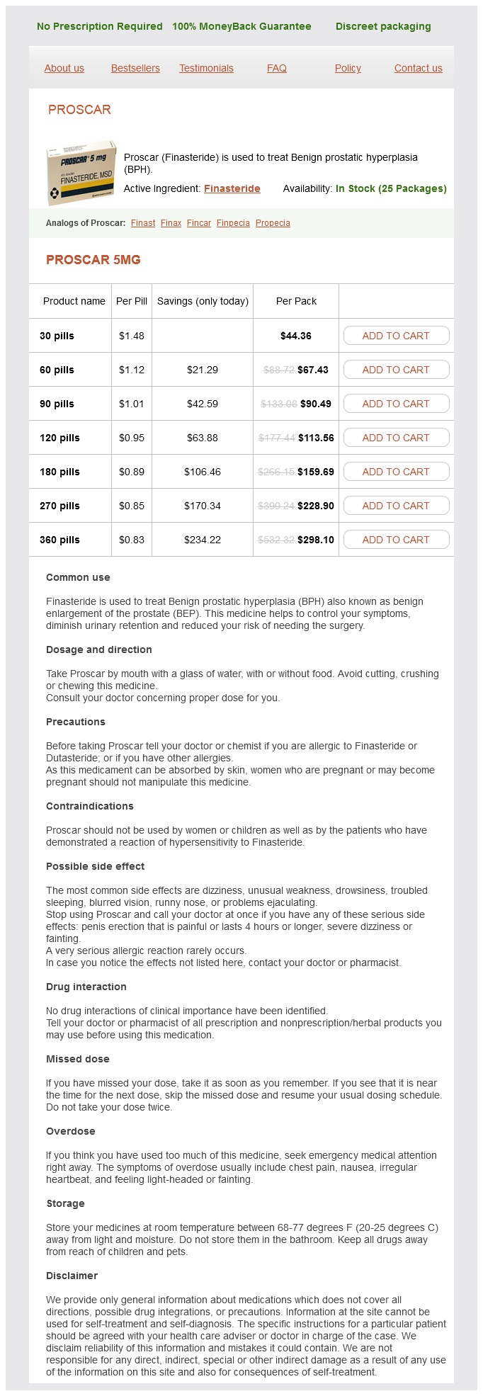
Proscar
Proscar
Proscar dosages: 5 mg
Proscar packs: 30 pills, 60 pills, 90 pills, 120 pills, 180 pills, 270 pills, 360 pills
In stock: 547
Only $0.88 per item
Description
The source of the pain is pressure in a contained space man health vitamin generic proscar 5 mg otc, pressing against nerve fibers and not from the soft tissue injury or bony injury alone. The nail matrix is the tissue under the base of the nail that permits nail growth and migration. Its longitudinal fibers anchor the dermis to the periosteum of the distal phalanx. Scarring of the matrix, as occurs with nail trauma, can disrupt nail growth and lead to nail deformity or permanent loss of the nail. When describing the injury, the clinician should estimate the percentage of nail bed that is affected by the hematoma and discuss associated trauma to the nail margins or surrounding tissue. The examination should include tests of the extensor and flexor tendons, of circulation by capillary refill, and of the sensitivity of the area. Crush injuries are associated with three types of distal phalanx fractures: longitudinal, transverse, and comminuted. If the fracture is angulated, displaced, unstable, or intraarticular or involves a third or more of the articular surface, refer the patient to a hand surgeon. In the two studies that examined this possible association,200,201 with fracture subsets totaling 26, no infections occurred. These studies suggest that the presence of an underlying fracture does not contraindicate nail trephination for fear of causing infection. There are no data supporting the routine use of prophylactic antibiotics following trephination if an underlying fracture is present. Subungual hematomas should be trephinated if they are acute (less than 24 to 48 hours old), are not spontaneously draining, are associated with intact nail folds, or are painful. After 48 hours, most subungual hematomas have clotted and trephination is typically not effective. Nail removal is unnecessary, even if there is an underlying distal phalanx fracture, as long as the nail margins and nail are intact. Simple trephination of an uncomplicated subungual hematoma, even if it involves the entire subungual area, results in a good outcome functionally and cosmetically. Prophylactic antibiotics are not necessary for patients with uncomplicated subungual hematomas that have been trephinated. For more serious injuries, a slight risk for infection exists, but in most cases rigorous wound cleaning and careful soft tissue repair constitute adequate treatment. Follow universal precautions because the fluid under the nail is under pressure and can spurt. Anesthesia is not always required, but a digital block can be used to calm an anxious patient and is commonly performed. An 18- or 21-gauge needle can be rotated between the thumb and the index finger while gentle pressure is applied so that the sharp end of the needle corkscrews through the nail. An alternative needle-based approach is being used by a group of Turkish dermatologists. Kaya and associates208 use extra fine, 29-gauge insulin syringes for evacuation of hematomas.
Resina Boswelliae (Indian Frankincense). Proscar.
- How does Indian Frankincense work?
- What is Indian Frankincense?
- Dosing considerations for Indian Frankincense.
- Are there safety concerns?
Source: http://www.rxlist.com/script/main/art.asp?articlekey=96109
Standard tubing is attached to the wall suction androgen hormone ovulation proscar 5 mg purchase visa, with the other end connected to the first suction canister. Each canister must have one port with self-sealing filter, which is needed to close the system. Once the suction canisters are connected, take the final suction tubing end and place into a syringe, with plunger removed. After the paracentesis catheter has been placed and flow obtained, attach the suction syringe to the catheter or first to a 3-way stopcock. Fluid should begin to drain into the first canister, and once full, fluid will continue into the other canisters in order. In addition, it allows the clinician to evaluate the most optimal location to attempt the procedure, which can be less straightforward in patients with smaller fluid collections or in whom physical examination alone is insufficient to make the diagnosis. Image Interpretation the location of ascites is variable and depends on the amount of fluid present, as well as the position of the patient. Evaluate the right upper quadrant by placing the transducer in either the transverse or the longitudinal orientation in the 8th to Continued Equipment Use a low-frequency transducer (2 to 5 mHz) to obtain a sufficient depth of penetration to visualize the area of interest. The transducer will probably need to be adjusted slightly to allow an optimal view of this area, either by moving up or down a rib space or by adjusting the angle of the transducer. Once this area has been evaluated in detail, evaluate the left upper quadrant in much the same fashion. Because the spleen is typically slightly more superior and posterior than the liver, place the transducer in the 8th to 11th intercostal space in the posterior axillary line. Ascitic fluid will appear as anechoic (black) areas in the splenorenal space, as well as the potential space between the diaphragm and the spleen. Once these areas have been evaluated, look at the pelvis and lower part of the abdomen by placing the transducer just superior to the pubic symphysis. The bladder is typically seen as a well-demarcated area that is rectangular in shape in the transverse orientation and slightly triangular in shape in the longitudinal orientation. In contrast, free fluid will appear to have irregular borders and may seem to "seep" into the crevices of the lower part of the abdomen and pelvis. The liver can be seen as the hypoechoic (dark gray) object on the left of the image, with anechoic (black) fluid seen between the liver and kidney (on the right of the image). Anechoic (black) free fluid can be seen surrounding the spleen at the right of the image. The diaphragm is seen as the bright white arcing structure on the far right of the image. Procedure and Technique Once free fluid has been seen, identify the area with the largest collection of fluid. Use ultrasound to identify the areas that contain the least number of loops of bowel. If the fluid collection appears to be large, use ultrasound to "mark the spot" and then put it aside. Proceed with the procedure in the typical sterile fashion in the area that has been identified and marked.
Specifications/Details
Tendons tend to not retract in this area mens health yoga 5 mg proscar purchase amex, so close inspection will usually result in location of the injured tendon. The decision to repair a partial tendon laceration in zone 4, and whether it should be repaired by the emergency provider, is best discussed with the consulting hand surgeon. In general, because of the duality of the extensor system in this region, lacerations of a single lateral slip can either be repaired with 5-0 nonabsorbable suture or left unrepaired and splinted. Minor lacerations in zone 4 that do not result in tension on the repair site can be treated with a finger guard for 7 to 10 days and early range of motion. Treat larger lacerations or those that result in tension at the repair site with a splint that extends from the forearm to the digit for 3 to 6 weeks. Group the fingers so that either the index and long fingers, or the long, ring, and little fingers are immobilized. Patients with wounds that are suspected of penetrating the joint are generally taken to the operating room for surgical exploration, irrigation, and treatment with intravenous antibiotics, but protocols vary. Zone 3 tendon lacerations can result in long-term deformity if not carefully repaired, and patients with such injuries are commonly referred to a hand surgeon. Partial lacerations of the central slip or lateral bands are managed variably, and it is advisable to discuss these injuries with the consulting hand surgeon. A boutonnière deformity develops when the central slip is ruptured by an open or closed mechanism that leads to unopposed action of the flexor digitorum superficialis tendon. Open central slip injuries are usually managed operatively, and complex injuries may require direct attachment of the tendon to bone or tendon reconstruction. Note the flexion of the proximal interphalangeal joint and extension of the distal interphalangeal joint from a laceration of the central slip mechanism. Promptly refer these patients to a hand surgeon so that repair may be undertaken within 1 week of injury. The tendon injury that is important to recognize in this setting is an occult isolated central slip rupture. With forced extension against resistance, patients usually have pain and may have decreased strength. In this position, a 15-degree or greater loss of active extension is highly suggestive of a central slip injury. To prevent the deformity from occurring, the provider should have a high index of suspicion for its presence and treat these patients conservatively. B, this splint allows active flexion at the metacarpophalangeal and distal interphalangeal joints. BE, the Elson test for early diagnosis of an acute rupture of the central slip of the extensor digitorum communis tendon. Such rupture results in a boutonnière deformity in which the distal interphalangeal joint is hyperextended, as shown.
Syndromes
- Endocarditis
- May get worse from drinking alcohol
- Fused fingers and toes
- Multiple myeloma
- Hypothalamic disease
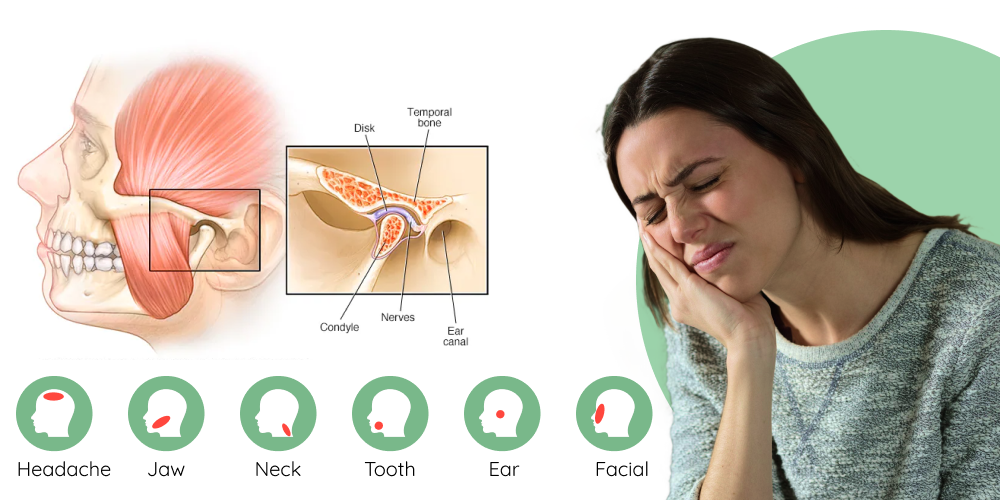
- Tooth decay
- Has it become worse over time?
- Weakness or drowsiness
Related Products
Additional information:
Usage: gtt.
Tags: generic proscar 5 mg with mastercard, proscar 5 mg order without prescription, buy proscar 5 mg lowest price, proscar 5 mg for sale
9 of 10
Votes: 93 votes
Total customer reviews: 93
Customer Reviews
Bram, 36 years: Digital examination reveals a large, tense, tender swelling along the anal canal that extends above the anorectal ring.
Tjalf, 28 years: A, Detachment of large sheets of necrolytic epidermis (>30% total body surface area) led to extensive areas of denuded skin.
Arakos, 33 years: Cakir B, Akan M, Yildirim S, et al: Localization and removal of ferromagnetic foreign bodies by magnet.
Arokkh, 57 years: Immobilizing tendon lacerations may facilitate the healing process by relieving stress on the repaired tendon.
Hauke, 47 years: Ideally, the emergency clinician should become familiar with a number of different techniques for reducing anterior shoulder dislocations because no single method has a 100% success rate nor is any technique ideal in every situation.
-
Our Address
-
For Appointment
Mob.: +91-9810648331
Mob.: +91-9810647331
Landline: 011 45047331
Landline: 011 45647331
info@clinicviva.in -
Opening Hours
-
Get Direction


