
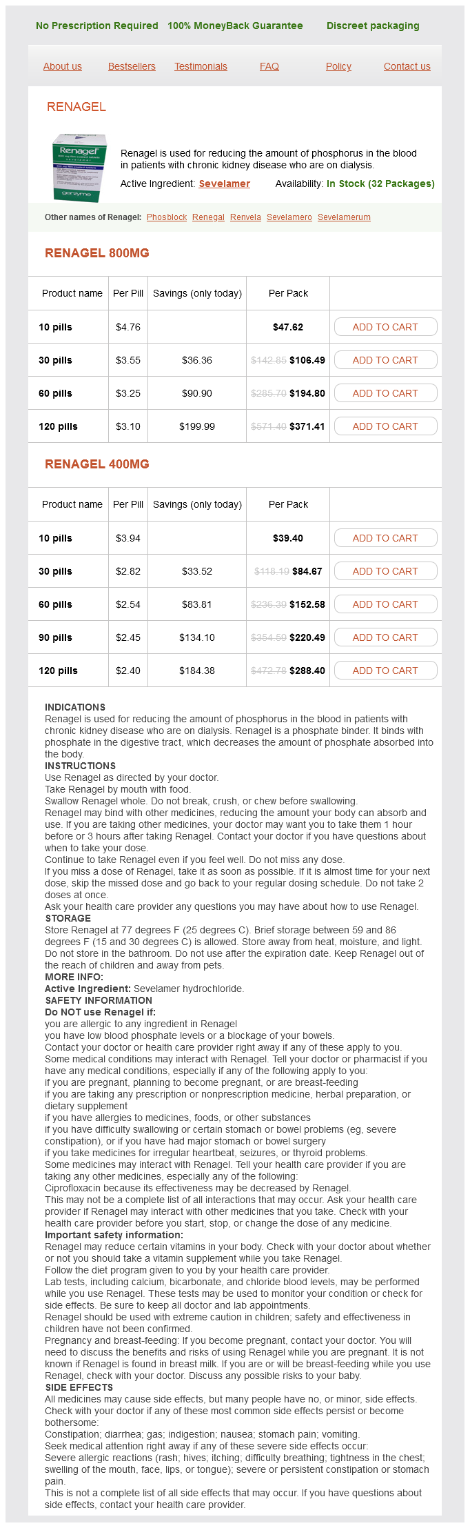
Renagel dosages: 800 mg, 400 mg
Renagel packs: 10 pills, 30 pills, 60 pills, 120 pills, 90 pills
In stock: 522
Only $2.55 per item
Once ossified gastritis diet ketosis renagel 400 mg order with mastercard, the tympanic cavity, mastoid antrum and the posterior end of the pharyngotympanic tube become surrounded by bone. The petrous part forms the roof, floor and medial wall of the cavity, while the squamous and tympanic parts, together with the tympanic membrane, form its lateral wall. At birth, the middle and inner ears are adult size, and the tympanic cavity, mastoid antrum, tympanic membrane and auditory ossicles are all almost adult size. The internal acoustic meatus is approximately 6 mm in horizontal diameter, 4 mm in vertical diameter and 7 mm in length at birth, and the adult diameters are 7. After birth and apart from general growth, the tympanic ring extends posterolaterally to become cylindrical, growing into a fibrocartilaginous tympanic plate, which forms the adjacent part of the external acoustic meatus at this stage. This growth is not equal but is rapid in the anterior and posterior regions, which meet and blend. Thus, for a time, an opening (foramen of Huschke) exists in the floor; it usually closes at about the fifth year but is sometimes permanent. The external acoustic meatus is relatively as long in children as it is in adults, but the canal is fibrocartilaginous, whereas its medial two-thirds are osseous in adults. Surgical access to the tympanic cavity is via the mastoid antrum, and in children it is necessary to remove only a thin scale of bone in the suprameatal triangle to reach the antrum. The tympanic plate ensheathes the styloid process by posterior extension, and extends medially over the petrous bone to the carotid canal. Initially, the mandibular fossa is shallow, facing more laterally, but it then deepens and ultimately faces downwards. Posteroinferiorly, the squamous part grows down behind the tympanic ring to form the lateral wall of the mastoid antrum. The mastoid part is at first flat, so that the stylomastoid foramen and rudimentary styloid process are immediately behind the tympanic ring. The lateral mastoid region grows downwards and forwards to form the mastoid process, which means that the styloid process and stylomastoid foramen become inferior. The mastoid process is not perceptible until late in the second year; consequently, the facial nerve is relatively superficial and susceptible to damage during surgical exploration during this period. In the neonate, the petrous and squamous parts of the temporal bone are usually partially separated by the petrosquamous fissure, which opens directly into the mastoid antrum of the middle ear. Rarely, the fissure closes in infants during the first year but it sometimes remains unclosed up to the age of 19 years; it is a route for the spread of infection from the middle ear to the meninges. There is a bimodal growth pattern in the lateral surface dimensions of the temporal bone in children; marked increases in dimension occur from birth to the age of 4 years but little growth is seen between the ages of 4 and 20 years (Simms and Neely 1989). The neonatal internal acoustic meatus is about half the length of its adult counterpart. Its opening from the middle ear cavity is as large as it is in the adult but the pharyngeal opening in the nasal part of the pharynx is relatively smaller.
Nimbaka (Lemon). Renagel.
Source: http://www.rxlist.com/script/main/art.asp?articlekey=96546
The base is concave gastritis diet ���� generic 800 mg renagel mastercard, with a smooth surface for articulation with the lateral part of the upper border of the cricoid lamina. Its round, prominent lateral angle, or muscular process, projects backwards and laterally; it gives attachment to posterior cricoarytenoid behind and lateral cricoarytenoid in front. The vocal ligament is attached to its pointed anterior angle (vocal process), which projects horizontally forwards. The apex curves backwards and medially, and articulates with the corniculate cartilage. It is smaller, but thicker and stronger, than the thyroid cartilage, and has a narrow curved anterior arch and a broad, flatter posterior lamina. They lie in the posterior parts of the aryepiglottic mucosal folds, and are sometimes fused with the arytenoid cartilages. Cricoid arch the cricoid arch is vertically narrow in front (57 mm in height) and widens posteriorly towards the lamina. Cricothyroid is attached to the external aspect of its front and sides, and cricopharyngeus (part of the inferior pharyngeal constrictor) is attached behind cricothyroid. The arch is palpable below the laryngeal prominence, from which it is separated by a depression containing the conus elasticus. The inferior border of the cartilage is nearly horizontal and is circular in outline, whereas the upper border is more elliptical. Their functions are unknown, although they may serve to strengthen this connection. They are the small, paired sesamoid cartilages, found at the lateral edges of the arytenoid cartilages, and a single interarytenoid cartilage, enclosed by the cricopharyngeal ligament. Each joint is enclosed by a capsular ligament and strengthened by a ligament that, although traditionally called the posterior cricoarytenoid ligament, is largely medial in position. The cricoid facets are elliptical, convex and obliquely directed laterally, anteriorly and downwards. The first is rotation of the arytenoid cartilages at right angles to the long axis of the cricoid facet (dorso-medio-cranial to ventrolatero-caudal), which, because of its obliquity, causes each vocal process to swing laterally or medially, thereby increasing or decreasing the width of the rima glottidis. This movement is sometimes referred to as a rocking movement of the arytenoid cartilages. There is also a gliding movement, by which the arytenoids approach or recede from one another, the direction and slope of their articular surfaces imposing a forward and downward movement on lateral gliding. When viewed from above, foreshortening can give the illusion that the arytenoid cartilages are rotating about their vertical axes, but the shape of the facets prevents such movement occurring (Selbie et al 1998). However, some authors maintain that rotatory movement about a vertical axis can occur (Liu et al 2013). The posterior cricoarytenoid ligaments limit forward movements of the arytenoid cartilages on the cricoid cartilage.
Upper and lower joint compartments the upper joint space contains approximately 1 gastritis pain remedy buy 400 mg renagel with amex. Both spaces may be considerably distended with fluid for diagnostic purposes in temporomandibular joint arthrography, for therapeutic purposes in arthrocentesis, and to improve access and visibility in arthroscopy. Both joint spaces have an anterior and posterior recess or pouch, and are lined by synovium, which has a grey hue when viewed through an arthroscope (see Video 32. The disc moves in an almost congruent manner relative to the condyle controlled by neuromuscular forces: the upper head of lateral pterygoid anteriorly and the elastic tissue in the bilaminar region posteriorly together pull the disc forwards or backwards. An alternative view is that a slippery articular disc doubles the number of virtually friction-free sliding surfaces, and so destabilizes the condyle in the same way that stepping on a banana skin destabilizes the foot. This bony partition is occasionally fenestrated by inflammatory processes or inadvertently during surgery to the joint, particularly for release of ankylosis, and rarely by the condyle being driven superiorly by violent trauma to the mandible. The maxillary artery and its proximal branches, most notably the middle meningeal artery, lie medially, just beyond the joint capsule. The uppermost part of the parotid capsule enclosing the branches of the facial nerve that supply the muscles of the upper face, including orbicularis oculi, are lateral to the joint capsule; the thread-like nerves are at risk during surgical approach to the joint. It is into this space anterior to the articular eminence that the condyle is displaced in cases of dislocation of the temporomandibular joint or fracture dislocation of the condylar head due to the vector of pull from lateral pterygoid, which inserts into the anterior condylar neck. Posteriorly, the tympanic plate and tegmen tympani separate the joint from the middle ear cavity. Perforation through this Functions of the articular disc the articulating surfaces of the mandibular condyle and the articular fossa are highly incongruent. Wear of the articular surfaces may be reduced by the presence of a deformable articular disc, which not only distributes compressive joint loading over a larger contact area but also significantly reduces the frictional force on the condyle and the articular fossa by separating slide and rotation into different joint compartments. The disc may aid joint lubrication by storing fluid squeezed out from loaded areas to create a weeping lubricant. In addition, its flexibility, due to its high water content and viscoelastic properties, allows it to function almost like a fluid, further reducing friction within the joint and thereby aiding joint lubrication. The friction between the articulating surfaces in the absence of the disc is 23 times greater than with the disc in situ. Removal of the disc increases surface friction within the joint by 23 times compared to joints with an intact disc. There is flattening, slight roughening and sclerosis of the condylar head and articular fossa. These changes, which are believed to be adaptive responses of the condyle and fossa to the absence of an intervening disc, collectively produce a broader contact, decreasing the load per unit area.
Syndromes
Additional information:
Usage: q.3h.
Tags: 800 mg renagel order, cheap renagel 800 mg without prescription, renagel 400 mg on-line, purchase renagel 800 mg line
Vak, 22 years: The latter two appear in the orbital plates between the fourth and fifth months in utero, and extend into the ethmoidal conchae. Scott J 1979 Qualitative and quantitative changes in the histology of the human submandibular salivary gland during postnatal growth. The contribution made by the frontonasal process to surface epithelial derivatives extends from the skin of the forehead, over the supraorbital and glabellar regions, including the upper eyelid and conjunctiva, to the external aspect of the nose and philtrum of the upper lip.
Hamlar, 65 years: Acting in concert, they could therefore assist the lateral rectus in abducting the visual axis, as in divergence of the eyes in transferring attention from near to far. They project backwards (curving posterolaterally) from the lateral ends of the body. This later subdivides into the blastema of superior, inferior, medial and lateral recti and inferior oblique.
Marlo, 56 years: Growth of a laryngocele is constrained by the surrounding tissues, and so it expands upwards into the paraglottic space anterior to the piriform fossa, and superiorly to expand the aryepiglottic fold and reach the vallecula (internal laryngocele). These approaches improve access to the nasal cavity, maxillary, ethmoidal and sphenoidal sinuses, the soft palate and nasopharynx, and the infratemporal fossa and pharyngeal space. The main symptom caused by infection of the pterygomandibular region is trismus (painful reflex muscle spasm), which usually affects medial pterygoid.
Knut, 39 years: The more cranially placed somites (at the lower right of the figure) are further developed than those caudally placed (at the upper left of the figure). The resulting variation in pupil area (maximally a factor of 16) will obviously affect the amount of light impinging on the retina. While crossing masseter, the duct lies between the upper and lower buccal branches of the facial nerve, and may receive the accessory parotid duct.
Oelk, 54 years: Discs are thinnest in the upper thoracic region and thickest in the lumbar region. The position of the fetal posterior fontanelle coincides with the junction between the superior angle of the squamous part of the occipital bone and the occipital angle of the parietal bone on either side. Its round, prominent lateral angle, or muscular process, projects backwards and laterally; it gives attachment to posterior cricoarytenoid behind and lateral cricoarytenoid in front.
C-585, Saraswati Vihar, Pitampura, New Delhi 110034


