
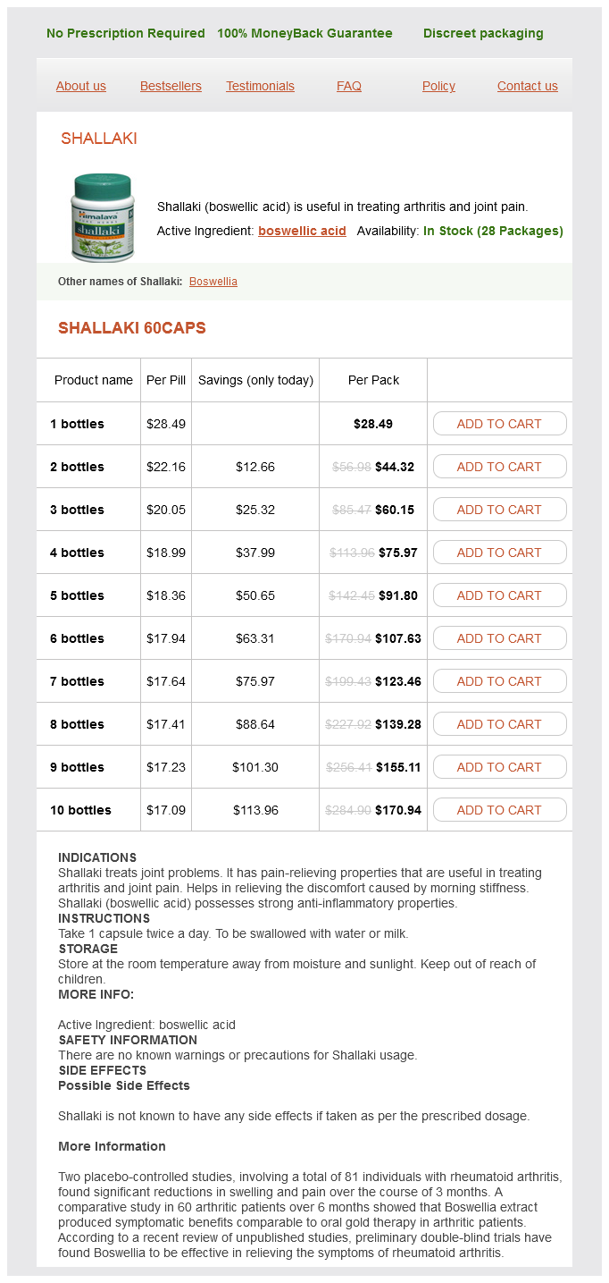
Shallaki dosages: 60 caps
Shallaki packs: 1 bottles, 2 bottles, 3 bottles, 4 bottles, 5 bottles, 6 bottles, 7 bottles, 8 bottles, 9 bottles, 10 bottles
In stock: 501
Only $18.16 per item
A small percentage of patients have a familial incidence of glomus tumors where the multiplicity rate may be as high as 30% muscle relaxant oil cheap shallaki 60 caps buy on-line. The displaced internal carotid artery flow void (white arrow), numerous serpentine flow voids in the lesion proper (black arrows), and "salt-n-pepper" appearance helps us a lot in the diagnosis of glomus tumor. B, Conventional catheter angiogram of the left common carotid in lateral projection (A, anterior; P, posterior) shows the avidly enhancing mass (M) displacing the internal (arrow) and external (arrowhead) arteries anteriorly, typical of glomus vagale. Perfect location for glomus vagale tumor (arrow): at C1 level, in carotid sheath, behind carotid artery and styloid musculature (arrowhead), enhancing nearly as much as vessels. When the carotid artery is encircled by tumor (arrow), you can bet that the surgeons will call the mass unresectable. Syndromic paragangliomas are also associated with succinate dehydrogenase germline mutations. There are rare reports that head and neck paragangliomas are linked to multiple endocrine neoplasia type 2 and von HippelLindau disease. Indium 111 octreotide scintigraphy enables distinction of glomus tumors from schwannomas and other masses of the carotid space as uptake occurs in the former but not the latter. False-positive cases can be seen in other neuroendocrine-like lesions such as medullary thyroid carcinomas, thyroid adenomas, Merkel cell tumors, and carcinoid lesions. Lymph nodes may be present anterior, posterior, medial, and lateral to the carotid sheath and therefore may displace the blood vessels in any possible direction. As a prequel to the brachial plexus section in this chapter, also noted on this image are the anterior scalene muscle (a) and the brachial plexus coursing posteriorly to this muscle (open arrows). B, Note the medial location of both internal carotid arteries (arrows) in the retropharyngeal space. Careful, spine surgeons, take note before you start your anterior approach spinal fusion! Often, one is called on to determine whether the carotid artery wall is invaded by spread from mucosal primaries or malignant lymphadenopathy. Mucosal Disease Spread the carotid artery and space may also be invaded from extension of mucosal cancers rather than lymphadenopathy. When a tumor surrounds the carotid sheath contents by over 270 degrees, carotid invasion is implied. Carotid blow-out in encased vessels figures into the therapeutic planning of the mucosal space squamous cell carcinoma. Radiation oncologists are generally reluctant (no guts, no glory) to radiate the bed of a tumor where the carotid artery may rupture. The incidence of rupture during radiotherapy is relatively small but is many times greater if the patient has already undergone surgery in the neck. Rarely, the interventionalist may occlude the diseased carotid artery before an attempt at complete surgical resection or radiotherapy. The retropharyngeal space extends from the base of the skull to the upper thoracic spinal level and is a site for spread from pharyngeal or esophageal lesions. Whereas the middle layer of the deep cervical fascia fuses with the deep layer at T6, the split in the deep layer may track to the level of the diaphragm.
Tuber Fleeceflower (Fo-Ti). Shallaki.
Source: http://www.rxlist.com/script/main/art.asp?articlekey=96750
The sequence of the numbers takes into account both the areas drained by the veins and the direction of blood flow iphone 5 spasms generic 60 caps shallaki fast delivery. This lateral view shows the internal cerebral vein (black arrowhead), vein of Galen (white arrow), straight sinus (black arrow), torcular (T), and superior sagittal sinus (S). Faintly seen are the basal vein of Rosenthal (r) and blush of the cavernous sinus (c). The internal cerebral veins are a marker for midline shift, behind the foramen of Monro. Deep Infratentorial Drainage the anatomy of posterior fossa venous drainage is more complex. Superior vermian, posterior pericallosal, mesencephalic, and internal occipital veins drain into the vein of Galen at the tentorial hiatus. The vein of Galen and the inferior sagittal sinus drain to the straight sinus, which in turn drains into the torcular herophili. From the torcular, blood flows to the transverse sinus, which receives drainage from the superior petrosal sinus, diploic veins, and lateral cerebellar veins. It courses laterally in the leaves of the tentorium and continues as the sigmoid sinus, which also drains the occipital sinus. The inferior vermian veins and superior hemispheric veins drain into the straight sinus. The transverse sinus receives blood from the inferior and superior hemispheric venous system. The deep middle cerebral vein courses deep in the sylvian fissure, and it meets with the anterior cerebral vein (which runs with the corresponding artery) to form the basal vein of Rosenthal arising along the brain stem. The basal vein of Rosenthal receives blood from the anterior pontomesencephalic vein in front of the brain stem, the lateral mesencephalic veins, and the precentral cerebellar vein (just anterior to the superior vermis behind the fourth ventricle). The black star represents the colliculocentral point, an angiographic landmark that should be about halfway between the tuberculum sellae and the torcular herophili. These venous channels receive blood from superior and inferior ophthalmic veins, the sphenoparietal sinus, and the superficial middle cerebral vein. The cavernous sinus communicates via (1) an extensive network across midline to the opposite cavernous sinus via the intercavernous sinus, (2) posteriorly via the superior petrosal sinus to the transverse sinuses just before the latter dive inferiorly to the sigmoid sinus, and (3) inferiorly into the inferior petrosal sinuses, which subsequently drain directly to the jugular bulbs. The inferior petrosal sinus also drains the internal auditory canal venous system. Arachnoid granulations often arise in the transverse and sigmoid venous sinuses and can simulate clots or other lesions. These granulations may also result in bony excavations in the adjacent skull, most commonly at the occipital skull but also along the frontal calvarium and sphenoid bone.
The caudate nucleus head indents the frontal horns of the lateral ventricle and is anterior to the globus pallidus; however muscle relaxant knots cheap 60 caps shallaki visa, the body of the caudate courses over the globus pallidus, paralleling the lateral ventricle and ending in a tail of tissue near the amygdala. The basal ganglia receive fibers from the sensorimotor cortex, thalamus, and substantia nigra, as well as from each other. The main function of the basal ganglia appears to be coordination of smooth movement. The other deep gray matter structures of interest in the supratentorial space are the thalami, which sit on either side of the third ventricle. The medial and lateral geniculate nuclei, located along the posterior aspect of the thalamus, serve as relay stations for visual and auditory function. In the infratentorial space, the dentate nucleus, the largest deep gray matter structure, has connections to the red nuclei and to the thalami. The archeocerebellum was separated and removed caudally from the middle cerebellar peduncle. Therefore, awareness of these cisterns and contents is critical in descriptions of different herniation syndromes and other pathologies that can be identified on imaging (Table 1-1). Meninges and Associated Potential Spaces the brain is covered in three protective layers of tissue, the meninges (or mater), which consist of pia, arachnoid, and dura. The dura (or pachymeninges) consists of the thickest and toughest layer and is adherent to the inner table of the skull extending to sutural margins. Pathologic conditions may occur in the space between the inner table and the dura, in the so-called epidural space, and result in mass effect on the underlying brain parenchyma. Epidural compartments are separated by sutures and pathologies typically do not cross the sutures when confined to the epidural space. Deep to the dura but superficial to the arachnoid mater is another potential space called the subdural space. The tiny dots of the fornix anteriorly (just ventral to asterisk) and the posterior commissure posteriorly (white arrow), as well as pulvinar thalamic gray natter (Pu), are also evident. The thalami are joined in the midline at the massa intermedia, and one can also see the forniceal columns (just below asterisk) projecting above the thalami. The chiasmatic recess (black arrow) and infundibular recess (black arrowhead) are also indicated. Note the crowding of structures at the foramen magnum in this patient with borderline Chiari I malformation. Subarachnoid disease processes occur at the surface of the brain, deep to the arachnoid layer but superficial to the pia, and therefore assume a curvilinear configuration, extending along the surface of the sulci and gyri. Subpial processes do occur, however, the pia and arachnoid cannot always be readily distinguished on imaging; the pia and arachnoid layers are collectively referred to as the leptomeninges. A small percentage (2% of children less than 8 years old and 10% of adolescents) of children show calcification of the pineal gland. Anterior to the pineal gland, one often sees the habenular commissure as a calcified curvilinear structure. The choroid plexus is calcified in about 5% of children by age 15, and most adults by age 40.
Syndromes
Additional information:
Usage: q.i.d.
Tags: order 60 caps shallaki with visa, shallaki 60 caps order mastercard, shallaki 60 caps without a prescription, generic 60 caps shallaki otc
Ayitos, 34 years: Wilson disease Wilson disease, described fully in Chapter 26, can produce an array of hyper- and hypokinetic movement disorders, including chorea, choreoathetosis, and dystonia. These are characteristic, with a slightly off-center spherical calcification of 1 to 2 mm in diameter, representing the scolex, surrounded by a partially or totally calcified sphere (7 to 12 mm in diameter).
Thorek, 51 years: Plaque erosion has an uncomplicated morphology with smooth borders on angiography. Nonetheless, a brief description of the speech pathway is warranted because of its critical role in humans.
Fasim, 60 years: In this population the average age at disease onset was 45 and the average period of survival was 27 years. The causes of orbital inflammatory disease include sinus infection (particularly in the pediatric population), bacteremia, skin infection (secondary to trauma, insect bite, and impetigo), or foreign body.
Aidan, 58 years: Serous choroidal detachment (benign prognosis) is crescentic or ring shaped, and the curvilinear choroid can be identified. They reported that although there was good intrafamilial similarity between patients there was interfamilial variation when patients of the same age were compared.
C-585, Saraswati Vihar, Pitampura, New Delhi 110034


