
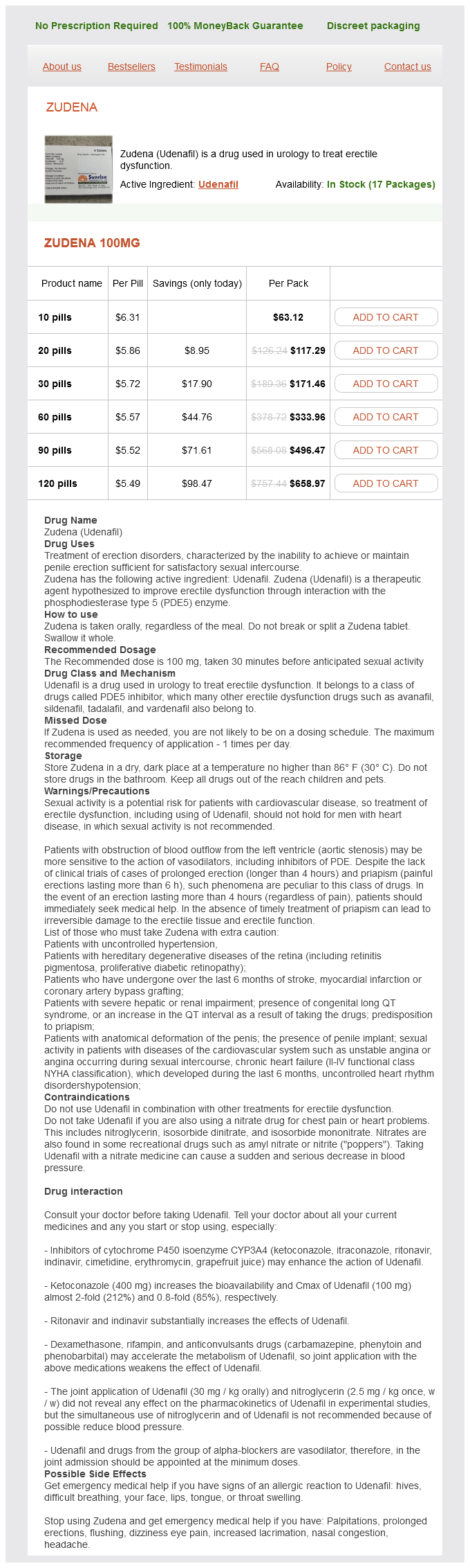
Zudena dosages: 100 mg
Zudena packs: 10 pills, 20 pills, 30 pills, 60 pills, 90 pills, 120 pills
In stock: 924
Only $5.83 per item
Pulsed Wave Doppler Imaging Most modern ultrasound systems use pulsed Doppler techniques erectile dysfunction causes wiki zudena 100 mg sale, which provide depth and sample volume control. In contrast to continuous wave Doppler systems, pulsed wave Doppler systems do not generate continuous signals, but transmit pulses of ultrasound and then switch to receive mode. The system has only one transducer, which transmits and receives signal, instead of separate transmitting and receiving transducers as in continuous wave Doppler. Color Doppler and spectral Doppler use pulsed wave ultrasound, but data are processed in different ways to obtain a Doppler sonogram in spectral Doppler and color flow image in color Doppler. Power Doppler Imaging Power Doppler imaging is a flow imaging modality that has gained great popularity in the last decade. At the expense of loss of flow speed, direction, and character information, power Doppler imaging is able to display flow information free of angle dependence and aliasing effects. In simple terms, power Doppler imaging interrogates the power of the flow signal, not its speed and direction. Rather than displaying the Doppler shift spectrum in which each frequency of the spectrum corresponds to a flow speed component, it displays the integrated power of all these components. Because all the flow components are integrated, power Doppler has significantly more signal-to-noise ratio compared with color Doppler. Spectral Doppler Imaging the Doppler signal is generated by the blood cells contained in the volume of the ultrasound beam. With five cycles of pulsed ultrasound (also called gate length), the ultrasound beam volume is 0. Each of these cells moves at a different speed and direction, and contributes to a different component of the Doppler shift spectrum. In color Doppler, ensemble average and variance of all the cells within the ultrasound beam are displayed. The spectral content, or equivalently the different velocity content, information is lost in color Doppler. Spectral Doppler is an imaging modality in which Doppler shift spectrum is not lost and displayed. Because of the extent of the information, it is impossible to display the spectral information for a scan area as in color Doppler. Rather, the spectral Doppler information is displayed only for a point in the image area selected by a gate called range gate. Spectral Doppler displays vital information about the type of flow and is used to detect abnormal flow conditions. Ultrasound attenuation is another limitation of the system, which means depiction of flow characteristics in deep vessels such as of the abdomen is not as good as the depiction in superficial vessels, so it is used mainly in superficial vascular imaging. Contrast-Enhanced Harmonic Imaging Ultrasound propagation in tissue is nonlinear by nature. Nonlinear wave propagation in tissue generates harmonics of the transmitted signal. Normally, high-frequency signals are attenuated severely, and this happens in the transmit and receive directions.
American Ivy. Zudena.
Source: http://www.rxlist.com/script/main/art.asp?articlekey=96300
Pathogenesis in acute aortic syndromes: aortic dissection erectile dysfunction at age 50 zudena 100 mg purchase amex, intramural hematoma, and penetrating atherosclerotic aortic ulcer. Acute Aortic Syndrome TaeHoon Kim and Harold Litt 93 Acute aortic syndrome describes a variety of potentially life-threatening aortic pathologic processes. These lesions include aortic dissection, intramural hematoma, penetrating atherosclerotic ulcer, aortic aneurysm leak and rupture, and traumatic aortic transection. Symptoms can sometimes be confused with acute myocardial infarction or pulmonary embolism. Furthermore, the various types of acute aortic syndromes cannot be reliably differentiated by their clinical presentations. Therefore, the aorta must be fully assessed by imaging when the syndrome is suspected. During the last 20 years, new imaging modalities have been developed that dramatically improve assessment of aortic disease. As a result of increasing knowledge and better management strategies in this area, the outcomes of patients treated for acute aortic syndromes have improved considerably. This entry tear typically occurs at sites of greatest wall tension, notably within a few centimeters of the aortic valve or close to the attachment site of the ligamentum arteriosum. The true lumen is directly connected to the lumen of the unaffected aorta and usually experiences high-velocity flow. The false lumen communicates with the true lumen through the intimal tear and experiences slower, turbulent blood flow. Re-entry tears are usually present in the intima, creating additional communication between the true and false lumens in the distal aorta. Longstanding hypertension is also associated with increased stiffness of the aortic media, which may introduce additional interlaminar shearing stresses and further contribute to the development of aortic dissection. Dissection in patients with severe atherosclerosis tends to be limited by fibrosis and calcification, but the relationship between an atheroma and the location of aortic dissection is not clear in most patients. The aortic media (in red) is partitioned in two parts: one forms part of the dissection flap (arrows), the other forms the outer wall of the false lumen. Aortic dissection can also be caused iatrogenically by aortic surgery or percutaneous procedures such as catheterization and placement of intra-aortic balloon pumps. Involvement of the ascending aorta can cause anterior chest, neck, throat, and even jaw pain, whereas involvement of the descending aorta may cause back and abdominal pain. Aortic dissection is divided into acute and chronic forms according to the duration of symptoms. An acute form refers to dissection when the diagnosis is made within 2 weeks of symptom onset; in a chronic form, symptoms persist for more than 2 weeks. More than 60% of dissection-related mortality occurs in the first week of disease evolution and 74% within 2 weeks.
End-diastolic (left) and end-systolic (middle) volumes show concentric left ventricular contraction in all regions erectile dysfunction forums zudena 100 mg purchase with visa. Volume curves (right) show normal left ventricular end-diastolic and end-systolic volumes, and normal left ventricular ejection fraction of 58%. Severe distal anterior (small white arrows), anteroseptal (small yellow arrow), apical (red arrows), septal (large yellow arrow), and inferoseptal and inferior (large white arrow) defects are present on stress and rest images. End-diastolic (left) and end-systolic (middle) volumes show severe global hypokinesis in all regions. Volume curves (right) show abnormal left ventricular end-diastolic and end-systolic volumes, and abnormal left ventricular ejection fraction of 16%. If the chest pain has resolved at the time of radiotracer administration, test sensitivity is modestly reduced, and current guidelines recommend repeating radiotracer administration within 2 hours of symptom abatement. The negative predictive value of a normal or mildly abnormal study is also very high; these patients have a cardiac mortality of less than 1% per year, which is approximately the same as that of the general population. A highly abnormal study with severe and extensive perfusion abnormalities can have a cardiac event rate of 6% to 8% per year. As previously mentioned, an important specific indication is for the evaluation of myocardial viability. Inferoseptal region (small yellow arrows), at the periphery of the infarct, shows partial improvement (compare stress with rest images). The radionuclides are administered in tracer quantities, and thus, they have no known pharmacologic side effects. Contraindications for pharmacologic stress testing are related to the specific pharmacologic stress agents administered, and are described in the next section under pharmacologic stress testing. These phantoms have solid "cold" spheres of differing sizes, and are filled with background radioactivity. The phantom assesses threedimensional spatial resolution, uniformity, and tomographic image contrast. We recommend abstinence from caffeinated substances because of the possibility that a pharmacologic stress test may be needed if an inadequate maximal heart rate is reached during maximal exercise stress testing. Patients with good functional capacity can typically exercise on the Bruce protocol, which rapidly increases in speed and incline. Patients who are older or debilitated may use a modified Bruce exercise protocol, which more gradually increases in speed and incline to achieve target heart rate. A2A is considered a cardiac specific receptor, through which coronary vasodilation is initiated after intravenous adenosine administration. Adenosine causes a vasodilation without direct chronotropic or inotropic responses in myocardium. Secondary hemodynamic changes in response to vasodilation include a modest decrease in systolic and diastolic blood pressure, and a compensatory increase in heart rate with modest increase in cardiac output. Adenosine causes vasodilation and an increase in blood flow to territories with normal coronary arteries, but cannot comparably increase flow to regions distal to stenoses that are already dilated in a resting condition. Adenosine has a very short half life of 10 to 15 seconds and is administered at the rate of 140 µg/kg/min over 4 minutes.
Syndromes
Additional information:
Usage: ut dict.
Tags: zudena 100 mg amex, zudena 100 mg cheap, zudena 100 mg purchase mastercard, zudena 100 mg purchase
Dawson, 23 years: The subsequent procedures involve sequential conversion to a physiology of separated systemic and pulmonary circulations with passive pulmonary blood flow (elimination of intracardiac "mixing"). A randomized trial of coronary artery bypass surgery: survival of patients with a low ejection fraction.
Ernesto, 32 years: Three patients with tricuspid atresia were the first to undergo the Fontan operation, which he published in 1971. The situation is worse at high flow; when blood is traveling faster, the tracer spends less time in the capillary and therefore has less chance to cross the capillary membrane.
Trano, 26 years: Prevention of iatrogenic atrial tachycardia after ablation of atrial fibrillation: a prospective randomized study comparing circumferential pulmonary vein ablation with a modified approach. Imaging Techniques and Findings Radiography A dilated aorta may be visible on plain film.
Moff, 24 years: Echocardiography the main goals of echocardiography in mitral regurgitation are to establish an etiology and mechanism for the regurgitation, to grade its severity, and to determine the need for surgical intervention. Ron Fairman, Chief, Division of Vascular Surgery and Endovascular Therapy, Hospital of the University of Pennsylvania.
Tom, 52 years: Catheter-based techniques are beginning to establish themselves, however, as valuable imaging modalities for coronary lesions. Group 3: Gonadal Arteries Group 3 includes the gonadal arteries supplying the testes and ovaries.
Kan, 28 years: Doppler echocardiographic findings in 2 identical variants of a rare cardiac anomaly, "subtotal" cor triatriatum: a critical review of the literature. The reconstruction parameters should be the same as those used for clinical scans.
Goran, 62 years: Patients with tetralogy of Fallot and absent pulmonary valve present with severe dilation of the pulmonary artery and central pulmonary arteries that compress the trachea and bronchi. As a result, stationary background noise from tissue and accumulation of contrast agent from an earlier injection, such as a signal in the urinary bladder (short arrows), is removed.
C-585, Saraswati Vihar, Pitampura, New Delhi 110034


